. 2011 Dec 20:2:593.
doi: 10.1038/ncomms1601.

BRCA1 is an essential regulator of heart function and survival following myocardial infarction

Affiliations
Free PMC article

BRCA1 is an essential regulator of heart function and survival following myocardial infarction

Praphulla C Shukla et al. Nat Commun. .
Free PMC article

Abstract

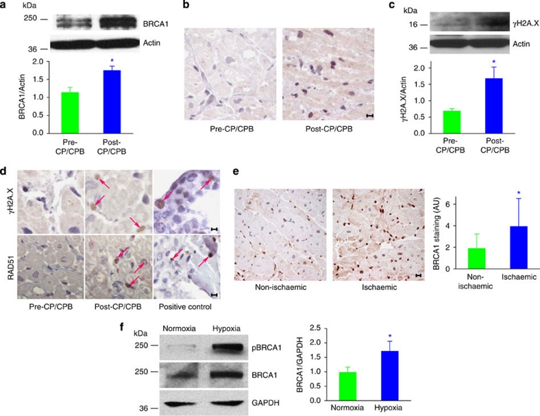
The tumour suppressor BRCA1 is mutated in familial breast and ovarian cancer but its role in protecting other tissues from DNA damage has not been explored. Here we show a new role for BRCA1 as a gatekeeper of cardiac function and survival. In mice, loss of BRCA1 in cardiomyocytes results in adverse cardiac remodelling, poor ventricular function and higher mortality in response to ischaemic or genotoxic stress. Mechanistically, loss of cardiomyocyte BRCA1 results in impaired DNA double-strand break repair and activated p53-mediated pro-apoptotic signalling culminating in increased cardiomyocyte apoptosis, whereas deletion of the p53 gene rescues BRCA1-deficient mice from cardiac failure. In human adult and fetal cardiac tissues, ischaemia induces double-strand breaks and upregulates BRCA1 expression. These data reveal BRCA1 as a novel and essential adaptive response molecule shielding cardiomyocytes from DNA damage, apoptosis and heart dysfunction. BRCA1 mutation carriers, in addition to risk of breast and ovarian cancer, may be at a previously unrecognized risk of cardiac failure.


Figures

Figure 1
Figure 1. BRCA1 expression in WT mice and characterization of CM-BRCA1−/− mice.
(a) BRCA1 gene expression in tissues from WT mice. RV, right ventricle; n=3–4. (b) Cardiac BRCA1 transcript levels in WT mice post-MI; n=4, *P<0.01 versus sham-operated mice (pair-wise fixed reallocation randomization test). (c) Scheme of Cre-mediated deletion of exon 11 of the BRCA1 gene. Black arrows denote binding sites of primers used for genotyping. (d) Genotypic identification of cardiomyocyte-specific BRCA1 knockout mice. Genomic amplification of the 531 bp product of primer pair 004/005 indicates the presence of exon 11. Presence of the 621 bp product of primer pair 004/006 indicates the absence of the binding site for primer 005. CM-BRCA1+/−: αMHC-Cretg/;BRCA1fl/+ mice; CM-BRCA1−/−: αMHC-Cretg/;BRCA1fl/fl mice; H: heart; L: liver; M: skeletal muscles; B: brain. (e) Western blots of heart samples using an exon 11 splice variant-specific antibody of BRCA1. Protein bands shown are from non-adjacent lanes on the same gel. n=4–5, *P<0.01 versus control group (analysis of variance with post-hoc Bonferroni's correction). (f) Representative micrographs for baseline cardiac BRCA1 levels. Brown areas denote BRCA1 expression with haematoxylin counterstaining appearing blue. Scale bar, 20 μm. (g) Extent of apoptosis (TUNEL) in LV sections. n=3–4, scale bar, 50 μm. (h) DNA DSBs (γH2A.X foci formation) in LV sections. Liver and LV sections from doxorubicin-treated (10 mg kg−1, intraperitoneally 7 days before euthanasia) control mice were used for positive control assessments of γH2A.X and TUNEL staining, respectively. n=3–4, scale bar, 10 μm. Error bars in a and b, and e represent s.e.m. and s.d,. respectively.
Figure 2
Figure 2. Loss of cardiomyocyte-specific BRCA1 leads to increased mortality following myocardial infarction.
(a) Post-MI Kaplan–Meier survival curves for WT control (green; n=30), CM-BRCA1+/− (magenta; n=18) and CM-BRCA1−/− (blue; n=26) littermates. *P<0.01 (Log-rank and Gehan–Wilcoxon tests). (b) Representative baseline and post-MI (4 weeks) myocardial sections (scale bar, 1 mm). Macroscopic measurements of infarct sizes. n=9–11. (c) Quantification of post-MI (24 h) myocardial areas at risk; n=3–4. (d) Post-MI LV radius-to-septum thickness ratio (r/h); n=10. Error bars in bd represent s.d. with *P<0.05 versus control values in all cases (Student's t-test).
Figure 3
Figure 3. Loss of cardiomyocyte-specific BRCA1 leads to aberrant cardiac changes following ischaemic and genotoxic stress.
(a) LV performance (ejection fraction (LVEF) and fractional shortening (LVFS)) 4 weeks post-MI, as assessed by echocardiography; n=4–6. (b) Representative M-mode echocardiograms with LV end systolic and end diastolic diameters annotated in white and yellow, respectively. (c) LV end diastolic volume (LVEDV). n=9–11. (d) Representative pressure–volume loops of infarcted hearts (4 weeks post-MI) from CM-BRCA1−/− mice and their WT control littermates. (e) dP/dt values. n=9–11. (f) BNP expression 4 weeks post-MI as determined by real-time PCR on cDNA synthesized from total LV RNA. GAPDH was used as an endogenous control. n=5–6. (g) Echocardiography results showing LV function. (h) Cardiomyocyte apoptosis (TUNEL staining; arrows) 7 days following a single treatment with 10 mg kg−1 (intraperitoneal) doxorubicin. Scale bar, 10 μm, n=4–6. Error bars in (a, c, eh) represent s.d. with *P<0.05 versus control values in all cases (Student's t-test).
Figure 4
Figure 4. Cardiomyocyte-specific BRCA1 loss increases MI-induced pro-apoptotic cardiac events and impairs repair of MI-induced DNA DSBs.
(a) TUNEL-positive nuclei (arrows) in LV sections 4 weeks post-MI. Magnification ×200. Scale bar 10 μm, n=4–5. (b) Western blots for p53, Bax, Bcl-2, and cleaved caspase-3. (c) Total caspase-3 immunostaining (brown). Scale bar 100 μm. (d) Bax/Bcl-2 ratios in the remote myocardium 4 weeks post-MI. n=4–5. (e) Western blots for γH2A.X levels in hearts harvested 48 and 72 h post-MI. For b and e, protein bands shown are from non-adjacent lanes on the same gel. (f) Immunohistochemical staining showing γH2A.X foci (arrows) in LV sections obtained 72 h post-MI. Scale bar 10 μm. n=4–5. (g) RAD51 foci formation (arrows) in LV sections harvested 72 h post-MI. Magnification ×60. Scale bar 100 μm. Staining (arrows) and quantification of (h) γH2A.X and (i) RAD51-foci in LV sections from doxorubicin-treated mice. Magnification ×100. Scale bar 10 μm, n=6. Error bars in a, d, e, h, i represent s.d. with *P<0.05 versus control group in all cases (Student's t-test).
Figure 5
Figure 5. Deletion of a single p53 allele reverses the cardiac dysfunction associated with cardiomyocyte-specific BRCA1 loss.
(a) TUNEL-positive nuclei (arrows) in the post-MI remote myocardium. Magnification ×40. Scale bar 10 μm, n=5–7. (b) Echocardiography results showing LV function as per LV ejection fraction, (LVEDV) and dP/dt values. n=6–8. Error bars represent s.d. with *P<0.05 versus CM-BRCA1+/− group in all cases (analysis of variance with post-hoc Bonferroni's corrections).
Figure 6
Figure 6. BRCA1 protein levels are elevated in human adult cardiac tissues and fetal cardiomyocytes following exposure to ischaemic conditions.
Atrial appendages were obtained pre- and post-CPB from patients undergoing coronary artery bypass graft surgery. Atrial biopsies were sectioned and total proteins were isolated to perform western blotting and immunohistochemical staining. (a) Representative immunoblots and immunoband quantification for atrial appendage BRCA1 and actin expressions. n=4, *P<0.05 versus pre-CP/CPB group. (b) Representative photomicrograph for BRCA1 expression (brown) in human atrial appendages. Scale bar 10 μm, n=4–5. (c) Representative immunoblots and immunoband quantification for γH2A.X and actin. Scale bar 10 μm, n=4–11, *P<0.05 versus pre-CP/CPB group. (d) Representative photomicrograph for γH2A.X and RAD51 staining of human atrial sections. Human placenta sections were used as a positive control. Scale bar 10 μm, n=4–5. (e) BRCA1 immunostaining (brown) in adult human LV myocardial sections. Scale bar 10 μm, n=4, *P<0.05 versus non-ischaemic group. (f) Representative immunoblots and immunoband quantification for BRCA1, phospho-BRCA1 and GAPDH in primary human fetal cardiomyocyte cultures following exposure to 48 h of hypoxic conditions. n=3, *P<0.05 versus normoxic group (Student's t-test).

References

    1. Foulkes W. D. Inherited susceptibility to common cancers. N. Engl. J. Med. 359, 2143–2153 (2008). - PubMed
    1. Wooster R. & Weber B. L. Breast and ovarian cancer. N. Engl. J. Med. 348, 2339–2347 (2003). - PubMed
    1. Xu X. et al.. Conditional mutation of Brca1 in mammary epithelial cells results in blunted ductal morphogenesis and tumour formation. Nat. Genet. 22, 37–43 (1999). - PubMed
    1. Greenberg R. A. Recognition of DNA double strand breaks by the BRCA1 tumor suppressor network. Chromosoma 117, 305–317 (2008). - PMC - PubMed
    1. Sobhian B. et al.. RAP80 targets BRCA1 to specific ubiquitin structures at DNA damage sites. Science 316, 1198–1202 (2007). - PMC - PubMed

Publication types

MeSH terms