. 2011 Dec 20:2:597.
doi: 10.1038/ncomms1594.

Promotion of plasma membrane repair by vitamin E

Affiliations
Free PMC article

Promotion of plasma membrane repair by vitamin E

Amber C Howard et al. Nat Commun. .
Free PMC article

Abstract

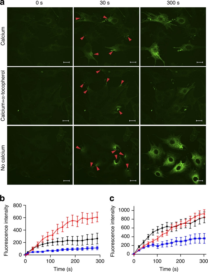
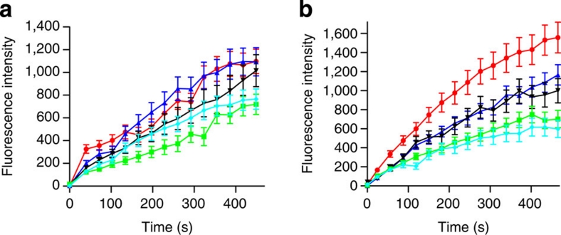
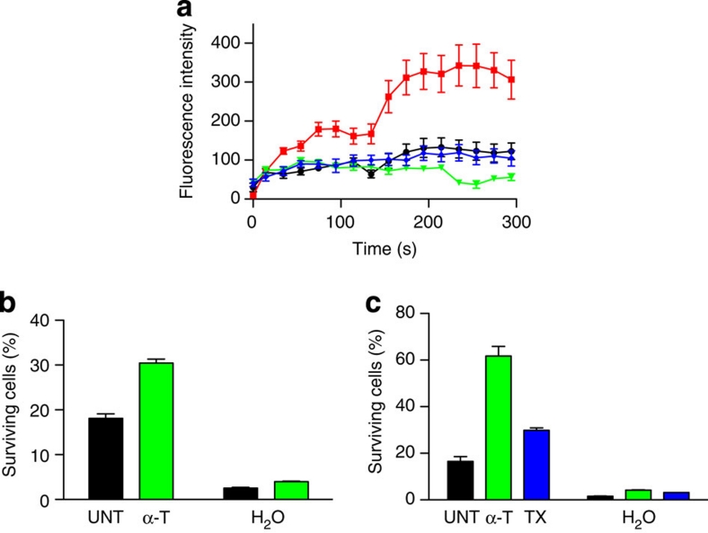
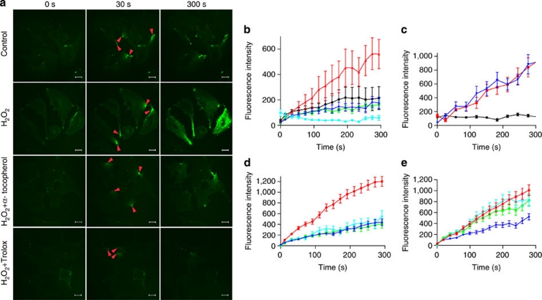
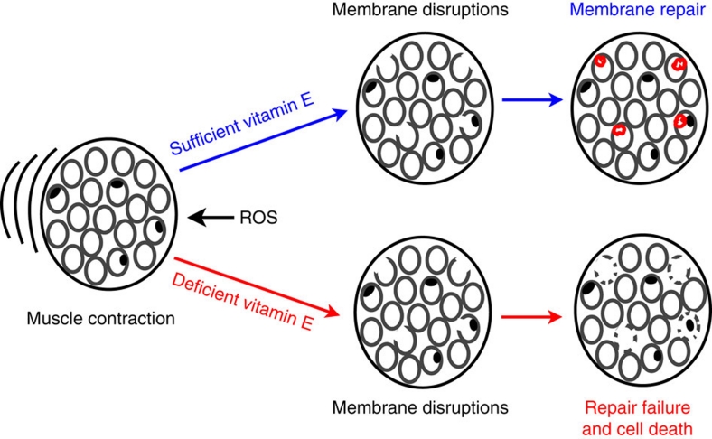
Severe vitamin E deficiency results in lethal myopathy in animal models. Membrane repair is an important myocyte response to plasma membrane disruption injury as when repair fails, myocytes die and muscular dystrophy ensues. Here we show that supplementation of cultured cells with α-tocopherol, the most common form of vitamin E, promotes plasma membrane repair. Conversely, in the absence of α-tocopherol supplementation, exposure of cultured cells to an oxidant challenge strikingly inhibits repair. Comparative measurements reveal that, to promote repair, an anti-oxidant must associate with membranes, as α-tocopherol does, or be capable of α-tocopherol regeneration. Finally, we show that myocytes in intact muscle cannot repair membranes when exposed to an oxidant challenge, but show enhanced repair when supplemented with vitamin E. Our work suggests a novel biological function for vitamin E in promoting myocyte plasma membrane repair. We propose that this function is essential for maintenance of skeletal muscle homeostasis.


Figures

Figure 1
Figure 1. Vitamin E promotes membrane repair in vitro.
(a) C2C12 cells were cultured for 18 h in a medium with or without 200 μM α-tocopherol, washed free of exogenous vitamin and immediately subjected to laser wounding in the presence or absence of calcium (added at 1.2 mM to PBS). Micrographs were taken before injury in FM 1-43 dye (time=0 s; the red arrowheads mark laser disruption sites) and imaged at 30 and 300 s after injury. (b) Quantitation of FM 1-43 dye influx over time after laser injury. C2C12 cells loaded with α-tocopherol (blue squares) before wounding displayed a significant (P<0.01; n=16) reduction in dye uptake after injury compared with non-treated controls (black dots); data for cells injured in the absence of added calcium is also presented (red triangles). (c) HeLa cells were loaded with 200 μM α-tocopherol for 24 h before laser analysis. Cells treated with α-tocopherol (blue squares) displayed a significant (P<0.001; n=15) reduction in dye uptake compared with control, untreated cells (black dots); data is also shown for cells treated with α-tocopherol and injured in the absence of added calcium (red triangles). Data are presented as the mean±s.e.m. Scale bars 20 μm.
Figure 2
Figure 2. Vitamin E loading interval and dosage.
(a) HeLa cells were loaded for 24 h with 2.5 (blue triangles), 25 (black triangles), 250 (green squares) and 2500 (cyan diamonds) μM α-tocopherol. No significant change in dye uptake compared with untreated controls (red dots) was found in cells treated with the lower concentrations of α-tocopherol. Only cells treated with 250 or 2500 μM α-tocopherol displayed a significant (P<0.001; n=21) decrease in dye uptake. (b) HeLa cells were loaded 48 h in the same concentrations as above. All samples treated with α-tocopherol (same colour scheme as 'b') displayed a significant (P<0.001; n=18) decrease in dye uptake relative to untreated controls (red dots). Data are presented as the mean±s.e.m.
Figure 3
Figure 3. Vitamin E reverses a high glucose-induced repair defect and enhances survival of mechanically injured cells.
(a) BS-C-1 cells were cultured in 30 mM glucose (red squares) for 14 weeks, a treatment previously shown to produce deficient membrane repair. Mannitol (30 mM) serves as an iso-osmolar control (black dots). Glucose-treated cells were loaded with 200 μM α-tocopherol (green triangles) or 200 μM Trolox (blue diamonds) for 24 h. Laser assay demonstrates a significant decrease (P<0.001; n=14) in dye uptake in the α-tocopherol (green triangles) and Trolox- (blue diamonds) treated cells compared with the untreated controls (red squares). (b) C2C12 myoblasts in a 96-well plate were loaded with 200 μM α-tocopherol (α-T; green bars) or left untreated (UNT; black bars) for 24 h. A live/dead assay was performed after mechanically scraping the cells off the plate in the presence of PBS containing Ca2+. Survival was calculated by reference to a non-wounded (not scraped) population of cells. Scraping of cells that were first osmotically shocked by immersion in distilled water provides a positive reference for cell death (H2O). A significant (P<0.0001; n=4) increase in survival was observed in the α-tocopherol-treated cells relative to untreated controls. (c) The same procedure, as in b, was followed for HeLa cells, and, in addition, a population of cells was treated with 200 μM water-soluble vitamin E (TX; blue bars). A significant (P<0.001; n=4) increase in survival was observed in the α-tocopherol and Trolox-treated cells relative to untreated controls. Data are presented as the mean±s.e.m.
Figure 4
Figure 4. Vitamin E prevents oxidative stress-induced repair failure.
(a) BS-C-1 cells were loaded for 24 h with 200 μM α-tocopherol or Trolox. Cells were laser wounded in PBS-containing calcium, in the presence or absence of 1 mM hydrogen peroxide (H2O2). Images of cells were captured before (0 s) and after injury (30 s and 300 s), red arrows indicate injury sites. (b) BS-C-1 cells not wounded but exposed to H2O2 (cyan dots), displayed nominal dye uptake in the laser assay. Cells wounded in the presence of H2O2 (red triangles) showed significantly (P<0.01; n=12) more dye influx than wounded cells not exposed to this oxidant challenge (black dots). Cell treatment with α-tocopherol (green squares) or Trolox (blue diamonds) significantly (P<0.01; n=17) decreased dye entry into oxidant-challenged cells. (c) BS-C-1 cells were laser wounded in the presence of 5 mM thimerosal (blue triangles). This oxidant significantly (P<0.01; n=12) increased dye uptake relative to untreated controls (black dots), to approximately the same level as cells injured in the absence of Ca2+ (red squares). (d) HeLa cells were pretreated 24 h with 200 μM α-tocopherol (green squares) or Trolox (blue diamonds), or 1 mM vitamin C (cyan triangles). Cells pretreated with antioxidants had significantly (P<0.05; n=17) less dye influx after laser injury than control, untreated cells (red dots). (e) HeLa cells were assessed for membrane repair immediately (less than 15 min) after addition of α-tocopherol (green squares), Trolox (blue diamonds), or vitamin C (cyan triangles) to the wounding solution. Only cells injured in Trolox displayed a significant (P<0.001; n=17) decrease in dye influx compared with untreated cells. Scale bars 20 μm.
Figure 5
Figure 5. Effect of antioxidants incapable of membrane association.
(a) HeLa cells were incubated for 24 h with 200 μM HRP (cyan triangles) or 200 μM α-tocopherol (green squares) or left untreated (black dots). Cells were washed free of exogenous antioxidants and laser wounded in the presence of calcium. One population of untreated cells was injured in PBS-containing HRP (magenta dots) or injured in the absence of calcium (red dots). Only cells that were loaded before wounding with α-tocopherol displayed a significant (P<0.05; n=17) decrease in dye uptake. (b) HeLa cells were injured in the presence or absence of 10 mM DTT (cyan triangles). Cells injured in the presence of DTT did not display a significant difference in dye uptake kinetics when compared with controls injured plus (black dots) or minus (red triangles) calcium. (c) BS-C-1 cells were injured in the presence of 10 mM DTT (cyan diamonds). Cells injured in the presence of DTT displayed no significant difference in dye uptake kinetics when compared with control untreated cells (black dots). The data representing wounding in the absence of Ca2+ is also shown (red triangles). Data are presented as the mean±s.e.m.
Figure 6
Figure 6. Vitamin E promotes membrane repair in intact skeletal muscle.
(a) Soleus muscle was excised and then with minimal delay laser injured (red arrow indicates the site of injury) in the presence of 5 mM hydrogen peroxide (H2O2). Wounding plus (calcium) and minus (no calcium) in the absence of H2O2 is also shown. One group of solei was treated with Trolox for 1 h before laser injury (Trolox). The confocal images depict myocyte staining before (0 s) and at two intervals (30 s and 445 s) after injury with FM 1-43 dye. (b) Measured FM dye influx (20 s intervals) for the four myocytes imaged above, injured in the absence of extracellular calcium (red triangles), in the presence of H2O2 (cyan squares), in the absence of H2O2 (black dots) and in the presence of H2O2 after Trolox pretreatment (magenta dots). Dips and rises in the fluorescence are observed due to muscle contractions that caused temporary lapses in focus. (c) Total dye uptake by myocytes in intact solei 445 s after laser injury under the conditions defined above. A significant (P<0.05; n=8) increase in dye uptake occurred in fibres exposed to H2O2 (blue bar) relative to unchallenged controls (red bar). Trolox pretreatment of H2O2 challenged cells (blue bar with diagonal stripes) significantly (P<0.05; n=10) decreased dye uptake compared with untreated H2O2 challenged cells (blue bar). Scale bars 20 μm.
Figure 7
Figure 7. A hypothesis for explaining how vitamin E promotes muscle homeostasis.
Muscle contractions, especially high-force eccentric contractions, disrupt the plasma membranes (grey ovals) of myocytes and generate ROS. If sufficient vitamin E is present in the membranes of these injured myocytes, repair succeeds via a patch mechanism (red circles in myocytes) and they survive this injury. If, on the other hand, the myocyte is deficient in vitamin E, repair fails, injured myocytes die and eventually myopathy develops.

References

    1. Bement W. M., Yu H. Y., Burkel B. M., Vaughan E. M. & Clark A. G. Rehabilitation and the single cell. Curr. Opin. Cell Biol. 19, 95–100 (2007). - PMC - PubMed
    1. Clarke M. S., Caldwell R. W., Chiao H., Miyake K. & McNeil P. L. Contraction-induced cell wounding and release of fibroblast growth factor in heart. Circ. Res. 76, 927–934 (1995). - PubMed
    1. McNeil P. L. & Khakee R. Disruptions of muscle fiber plasma membranes. Role in exercise-induced damage. Am. J. Pathol. 140, 1097–1109 (1992). - PMC - PubMed
    1. McNeil P. L. & Ito S. Gastrointestinal cell plasma membrane wounding and resealing in vivo. Gastroenterology 96, 1238–1248 (1989). - PubMed
    1. McNeil P. L. & Ito S. Molecular traffic through plasma membrane disruptions of cells in vivo. J. Cell Sci. 96, 549–556 (1990). - PubMed

Publication types

MeSH terms