. 2012 Feb 10;287(7):5112-21.
doi: 10.1074/jbc.M111.306316. Epub 2011 Dec 20.

Characterization of SfmD as a Heme peroxidase that catalyzes the regioselective hydroxylation of 3-methyltyrosine to 3-hydroxy-5-methyltyrosine in saframycin A biosynthesis

Affiliations

Characterization of SfmD as a Heme peroxidase that catalyzes the regioselective hydroxylation of 3-methyltyrosine to 3-hydroxy-5-methyltyrosine in saframycin A biosynthesis

Man-Cheng Tang et al. J Biol Chem. .

Abstract

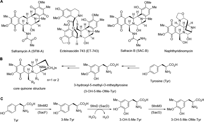
Saframycin A (SFM-A) is a potent antitumor antibiotic that belongs to the tetrahydroisoquinoline family. Biosynthetic studies have revealed that its unique pentacyclic core structure is derived from alanine, glycine, and non-proteinogenic amino acid 3-hydroxy-5-methyl-O-methyltyrosine (3-OH-5-Me-OMe-Tyr). SfmD, a hypothetical protein in the biosynthetic pathway of SFM-A, was hypothesized to be responsible for the generation of the 3-hydroxy group of 3-OH-5-Me-OMe-Tyr based on previously heterologous expression results. We now report the in vitro characterization of SfmD as a novel heme-containing peroxidase that catalyzes the hydroxylation of 3-methyltyrosine to 3-hydroxy-5-methyltyrosine using hydrogen peroxide as the oxidant. In addition, we elucidated the biosynthetic pathway of 3-OH-5-Me-OMe-Tyr by kinetic studies of SfmD in combination with biochemical assays of SfmM2, a methyltransferase within the same pathway. Furthermore, SacD, a counterpart of SfmD involved in safracin B biosynthesis, was also characterized as a heme-containing peroxidase, suggesting that SfmD-like heme-containing peroxidases may be commonly involved in the biosynthesis of SFM-A and its analogs. Finally, we found that the conserved motif HXXXC is crucial for heme binding using comparative UV-Vis and Magnetic Circular Dichroism (MCD) spectra studies of SfmD wild-type and mutants. Together, these findings expand the category of heme-containing peroxidases and set the stage for further mechanistic studies. In addition, this study has critical implications for delineating the biosynthetic pathway of other related tetrahydroisoquinoline family members.


Figures

FIGURE 1.
FIGURE 1.
Structures of representative tetrahydroisoquinoline family members with their common features and the proposed biosynthetic pathway of 3-OH-5Me-OMe-Tyr. A, structures of the representative members of tetrahydroisoquinoline family natural products. B, core quinone structure of these members and its deduced biosynthetic precursors. C, proposed biosynthetic pathway of 3-OH-5-Me-OMe-Tyr involved in SFM-A or SAC-B biosynthesis.
FIGURE 2.
FIGURE 2.
Protein purification and cofactor identification of SfmD. A, purification of recombinant C-His8-tagged SfmD as monitored by SDS-PAGE. Lane M, molecular weight marker; lane 1, total proteins after IPTG induction; lane 2, total soluble proteins; lane 3, purified protein. B, UV-Vis spectra of SfmD. SfmD without (i) and with (ii) addition of ALA when the protein was induced to express. C, characterization of SfmD by LC-MS. SfmD without (i) and with (ii) addition of ALA during the protein expression. D, pyridine hemichrome and hemochrome assays of SfmD, the amount of heme bound to SfmD was calculated by following the absorbance change at 553 nm using a difference extinction coefficient of 23.76 mm−1 cm−1 (17). (i) the hemochrome of SfmD; (ii) the hemichrome of SfmD.
FIGURE 3.
FIGURE 3.
Characterization of SfmD as a heme-containing peroxidase. A, H2O2 dismutation experiments recorded in UV-Vis spectra at 240 nm. The solution of H2O2 in the absence (i) or presence (ii) of SfmD (25 μm). B, low temperature EPR spectra of SfmD (50 μm). Spectra of SfmD resting state (top) indicating the high-spin ferric signals (g = 2.0 and g = 5.9) and its peroxide-activated form (Compound I) (bottom). C, HPLC analysis of the hydroxylation of 3-Me-Tyr to 3-OH-5-Me-Tyr catalyzed by SfmD: authentic standard of 3-OH-5-Me-Tyr (i) and 3-Me-Tyr (ii), reaction with O2 (iii) and H2O2 (iv) as the oxidant, negative control with boiled enzyme (v); (●), 3-OH-5-Me-Tyr, (ο), 3-Me-Tyr. D, LC-MS analysis of the production of 3-OH-5-Me-Tyr using H2O2 (i) and H218O2 (ii) as the oxidant.
FIGURE 4.
FIGURE 4.
Kinetic analysis of the hydroxylation reaction catalyzed by SfmD with substrate 3-Me-Tyr (A) or Tyr (B). The reaction mixtures consist of 50 μm protein, excess H2O2 (4 mm) and variable substrate (25 μm-2 mm) under optimized conditions.
FIGURE 5.
FIGURE 5.
UV-Vis spectra of SacD (A) and HPLC analysis of the hydroxylation of 3-Me-Tyr to 3-OH-5-Me-Tyr catalyzed by SacD (B). (i) Authentic standard of 3-Me-Tyr; (ii) authentic standard of 3-OH-5-Me-Tyr; (iii) negative control with boiled enzyme; (iv) reaction mixture; (●), 3-OH-5-Me-Tyr, (○), 3-Me-Tyr.
FIGURE 6.
FIGURE 6.
Multiple sequence alignment of SfmD and its analogs. Amino acid sequences were obtained from GenBankTM, including: SfmD from Streptomyces lavendulae NRRL 11002; SacD from Pseudomonas fluorescens A2–2; AZL_f00720 from Azospirillum sp. B510; KSE_08540 from Kitasatospora setae KM-6054.
FIGURE 7.
FIGURE 7.
UV-Vis spectra (A) and MCD spectra (B) of SfmD wild type and mutants. SfmD WT (black solid), SfmD H191A (purple dot), SfmD H274A (green dot), SfmD H313A (red dot), SfmD C317A (blue dash).

Similar articles

Cited by

References

    1. Scott J. D., Williams R. M. (2002) Chemistry and biology of the tetrahydroisoquinoline antitumor antibiotics. Chem. Rev. 102, 1669–1730 - PubMed
    1. Arai T., Takahashi K., Kubo A. (1977) New antibiotics saframycins A, B, C, D, and E. J. Antibiot. 30, 1015–1018 - PubMed
    1. Cuevas C., Francesch A. (2009) Development of Yondelis (trabectedin, ET-743). A semisynthetic process solves the supply problem. Nat. Prod. Rep. 26, 322–337 - PubMed
    1. Mikami Y., Takahashi K., Yazawa K., Arai T., Namikoshi M., Iwasaki S., Okuda S. (1985) Biosynthetic studies on saframycin A, a quinone antitumor antibiotic produced by Streptomyces lavendulae. J. Biol. Chem. 260, 344–348 - PubMed
    1. Palaniswamy V. A., Gould S. J. (1986) The incorporation of 3′-methyltyrosine and 5′-methyl DOPA into naphthyridinomycin. J. Am. Chem. Soc. 108, 5651–5652

Publication types

MeSH terms

LinkOut - more resources