. 2012 Feb 17;287(8):6002-13.
doi: 10.1074/jbc.M111.310110. Epub 2011 Dec 20.

Up-regulation of Cavβ3 subunit in primary sensory neurons increases voltage-activated Ca2+ channel activity and nociceptive input in neuropathic pain

Affiliations

Up-regulation of Cavβ3 subunit in primary sensory neurons increases voltage-activated Ca2+ channel activity and nociceptive input in neuropathic pain

Li Li et al. J Biol Chem. .

Abstract

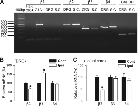
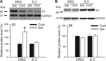
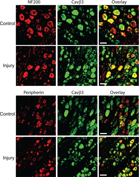
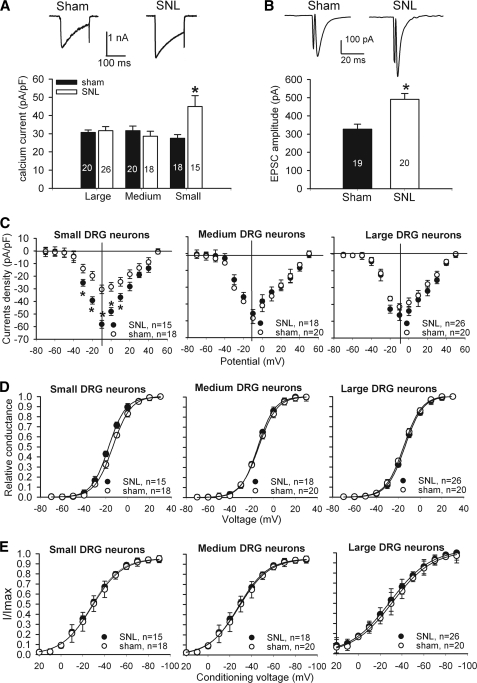
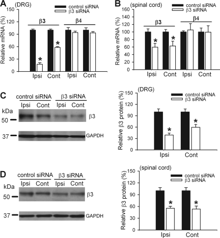
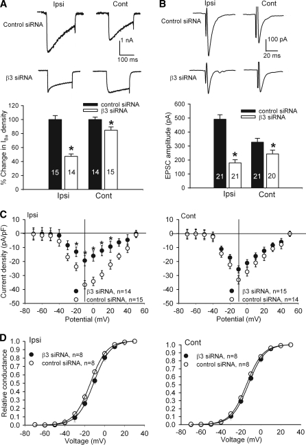
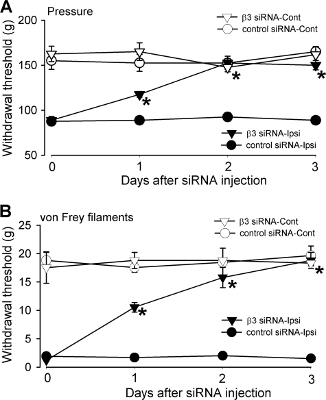
High voltage-activated calcium channels (HVACCs) are essential for synaptic and nociceptive transmission. Although blocking HVACCs can effectively reduce pain, this treatment strategy is associated with intolerable adverse effects. Neuronal HVACCs are typically composed of α(1), β (Cavβ), and α(2)δ subunits. The Cavβ subunit plays a crucial role in the membrane expression and gating properties of the pore-forming α(1) subunit. However, little is known about how nerve injury affects the expression and function of Cavβ subunits in primary sensory neurons. In this study, we found that Cavβ(3) and Cavβ(4) are the most prominent subtypes expressed in the rat dorsal root ganglion (DRG) and dorsal spinal cord. Spinal nerve ligation (SNL) in rats significantly increased mRNA and protein levels of the Cavβ(3), but not Cavβ(4), subunit in the DRG. SNL also significantly increased HVACC currents in small DRG neurons and monosynaptic excitatory postsynaptic currents of spinal dorsal horn neurons evoked from the dorsal root. Intrathecal injection of Cavβ(3)-specific siRNA significantly reduced HVACC currents in small DRG neurons and the amplitude of monosynaptic excitatory postsynaptic currents of dorsal horn neurons in SNL rats. Furthermore, intrathecal treatment with Cavβ(3)-specific siRNA normalized mechanical hyperalgesia and tactile allodynia caused by SNL but had no significant effect on the normal nociceptive threshold. Our findings provide novel evidence that increased expression of the Cavβ(3) subunit augments HVACC activity in primary sensory neurons and nociceptive input to dorsal horn neurons in neuropathic pain. Targeting the Cavβ(3) subunit at the spinal level represents an effective strategy for treating neuropathic pain.


Figures

FIGURE 1.
FIGURE 1.
Effect of nerve injury on the mRNA level of Cavβ subunits in the DRG and dorsal spinal cord tissues. A, agarose gel electrophoresis showing the presence of the DNA products of Cavβ2, Cavβ3, and Cavβ4 subunits in the DRG and dorsal spinal cord (S.C.) tissues from control rats. G1A1 cells stably expressing Cavβ1 were used as a positive control for the Cavβ1 subunit, and the HEK293A cell line was used as a negative control. B, group data show the effect of SNL on the mRNA level of Cavβ2, Cavβ3, and Cavβ4 subunits in the DRG (n = 4 in each group). C, summary data show the effect of SNL on the mRNA level of Cavβ2, Cavβ3, and Cavβ4 subunits in the dorsal spinal cord (SC) (n = 4 in each group). Ipsi, ipsilateral (nerve injury) side; Cont, contralateral (control) side. *, p < 0.05, compared with the corresponding value on the contralateral (control) side.
FIGURE 2.
FIGURE 2.
Effect of nerve injury on the protein level of Cavβ3 and Cavβ4 subunits in the DRG and dorsal spinal cord tissues. A, Western blots (top) and quantification (bottom) of the protein amount of Cavβ3 and Cavβ4 subunits in the DRG ipsilateral and contralateral to SNL (n = 4 in each group). B, Western blots (top) and quantification (bottom) of the protein amount of Cavβ3 and Cavβ4 subunits in the dorsal spinal cord (SC) ipsilateral and contralateral to SNL (n = 4 in each group). Ipsi, ipsilateral (nerve injury) side; Cont, contralateral (control) side. *, p < 0.05, compared with the corresponding value on the contralateral (control) side.
FIGURE 3.
FIGURE 3.
Nerve injury induces increased Cavβ3 in peripherin-immunoreactive DRG neurons. Representative confocal images show the distribution of Cavβ3 immunoreactivity (green) in small and large DRG neurons obtained from a sham control and an SNL rat. Neurons that are immunoreactive to NF200 or peripherin are indicated in red. Scale bar, 50 μm. All images are single confocal optical sections.
FIGURE 4.
FIGURE 4.
Effects of nerve injury on HVACC activity of DRG neurons and monosynaptic EPSCs of spinal dorsal horn neurons. A, original current traces (upper panel) show HVACC currents in small DRG neurons from a control and a nerve-injured rat. Group data (lower panel) show that SNL increased HVACC currents in small, but not large and medium, DRG neurons. Neurons were voltage-clamped at −90 mV and depolarized to 0 mV for 200 ms. The number of cells in each group is indicated in the column. B, representative traces and group data show the monosynaptic EPSCs of lamina II neurons in spinal cords obtained from sham control (n = 19) and SNL (n = 20) rats. C, effect of SNL on the current-voltage relationship of HVACCs in small, medium, and large DRG neurons. D, effect of SNL on voltage-dependent activation of HVACCs in small, medium, and large DRG neurons. Note that SNL shifted voltage-dependent activation of HVACCs to the left only in small DRG neurons. The V0.5 in control and SNL rats was −13.7 ± 0.1 and −17.5 ± 0.1 mV (p < 0.05), respectively. The slope factor in control and SNL rats was 8.33 ± 0.2 and 7.80 ± 0.1 mV (p > 0.05), respectively. E, effect of SNL on voltage-dependent inactivation of HVACCs in small, medium, and large DRG neurons. *, p < 0.05, compared with the corresponding value in the sham group. pF, picofarad.
FIGURE 5.
FIGURE 5.
Effect of intrathecal treatment with Cavβ3-specific siRNA (5 μg per day for 3 days) on the expression level of Cavβ3 in the DRG and spinal cord of SNL rats. A, effect of Cavβ3-specific siRNA on the mRNA level of Cavβ3 and Cavβ4 in the DRG of SNL rats (n = 4 in each group). B, effect of Cavβ3-specific siRNA on the mRNA level of Cavβ3 and Cavβ4 in the dorsal spinal cord of SNL rats (n = 4 in each group). C, Western blots (left) and quantification (right, n = 4 in each group) of the protein amount of the Cavβ3 subunit in the DRG ipsilateral and contralateral to SNL in rats treated with Cavβ3-specific siRNA or control siRNA. D, Western blots (left) and quantification (right, n = 4 in each group) of the protein amount of the Cavβ3 subunit in the dorsal spinal cord ipsilateral and contralateral to SNL in rats treated with Cavβ3-specific siRNA or control siRNA. Ipsi, ipsilateral (nerve injury) side; Cont, contralateral (control) side. *, p < 0.05, compared with the corresponding value in control siRNA-treated group.
FIGURE 6.
FIGURE 6.
Effects of intrathecal treatment with Cavβ3-specific siRNA on HVACC activity in DRG neurons and monosynaptic EPSCs of spinal dorsal horn neurons of SNL rats. A, original traces and group data show the effect of Cavβ3-specific siRNA on HVACC current in small DRG neurons ipsilateral and contralateral to SNL. The number of cells in each group is indicated in the column. B, representative traces and summary data show the effect of Cavβ3-specific siRNA on the amplitude of monosynaptic EPSCs of lamina II neurons in the spinal cord of SNL rats. C, effect of Cavβ3-specific siRNA on the current-voltage relationship of HVACCs in small DRG neurons ipsilateral and contralateral to SNL. D, effect of Cavβ3-specific siRNA on voltage-dependent activation of HVACCs in small DRG neurons ipsilateral and contralateral to SNL. Note that treatment with Cavβ3-specific siRNA shifted the voltage-dependent activation of HVACCs to the right in small DRG neurons ipsilateral to SNL. The V0.5 in control-siRNA and Cavβ3-specific siRNA groups was −14.37 ± 0.1 and −11.02 ± 0.1 mV (p < 0.05), respectively. The slope factor in control-siRNA and Cavβ3-specific siRNA groups was 8.20 ± 0.1 and 8.20 ± 0.2 mV (p > 0.05), respectively. Ipsi, ipsilateral (nerve injury) side; Cont, contralateral (control) side. *, p < 0.05, compared with the corresponding value in control siRNA-treated group.
FIGURE 7.
FIGURE 7.
Effects of intrathecal treatment with Cavβ3-specific siRNA (5 μg per day for 3 days) on nociceptive and tactile withdrawal thresholds of hind paws in SNL rats. A, time course effects of control siRNA and Cavβ3-specific siRNA on the nociceptive withdrawal threshold, tested with a noxious pressure stimulus, of hind paws ipsilateral and contralateral to SNL (n = 6 rats in each group). B, time course effects of control siRNA and Cavβ3-specific siRNA on the tactile withdrawal threshold, measured with von Frey filaments, of hind paws ipsilateral and contralateral to SNL (n = 6 rats in each group). Note that Cavβ3-specific siRNA, but not control siRNA, increased nociceptive and tactile withdrawal thresholds of hind paws ipsilateral to SNL. Ipsi, ipsilateral (nerve injury) side; Cont, contralateral (control) side. *, p < 0.05, compared with the base-line value in Cavβ3-specific siRNA-treated group.

Similar articles

Cited by

References

    1. Dworkin R. H., O'Connor A. B., Backonja M., Farrar J. T., Finnerup N. B., Jensen T. S., Kalso E. A., Loeser J. D., Miaskowski C., Nurmikko T. J., Portenoy R. K., Rice A. S., Stacey B. R., Treede R. D., Turk D. C., Wallace M. S. (2007) Pharmacologic management of neuropathic pain. Evidence-based recommendations. Pain 132, 237–251 - PubMed
    1. Campero M., Serra J., Marchettini P., Ochoa J. L. (1998) Ectopic impulse generation and autoexcitation in single myelinated afferent fibers in patients with peripheral neuropathy and positive sensory symptoms. Muscle Nerve 21, 1661–1667 - PubMed
    1. Gracely R. H., Lynch S. A., Bennett G. J. (1992) Painful neuropathy. Altered central processing maintained dynamically by peripheral input. Pain 51, 175–194 - PubMed
    1. Catterall W. A., Few A. P. (2008) Calcium channel regulation and presynaptic plasticity. Neuron 59, 882–901 - PubMed
    1. Catterall W. A. (2000) Structure and regulation of voltage-gated Ca2+ channels. Annu. Rev. Cell Dev. Biol. 16, 521–555 - PubMed

Publication types

MeSH terms

LinkOut - more resources