Review
. 2011 Dec;4(6):711.
doi: 10.1161/CIRCGENETICS.110.957829.

Comprehensive analysis of protein modifications by top-down mass spectrometry

Affiliations
Review

Comprehensive analysis of protein modifications by top-down mass spectrometry

Han Zhang et al. Circ Cardiovasc Genet. 2011 Dec.

Abstract

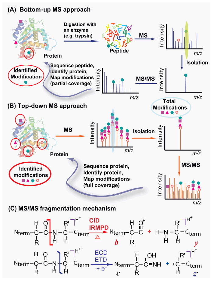
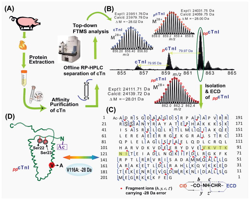
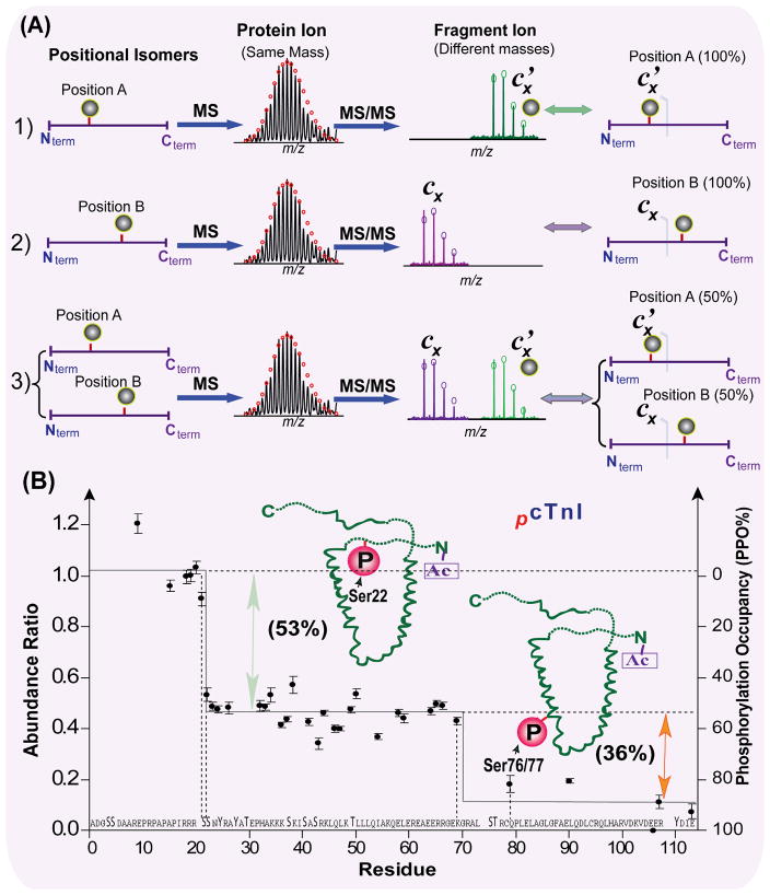
Mass spectrometry (MS)-based proteomics is playing an increasingly important role in cardiovascular research. Proteomics includes identification and quantification of proteins and the characterization of protein modifications, such as posttranslational modifications and sequence variants. The conventional bottom-up approach, involving proteolytic digestion of proteins into small peptides before MS analysis, is routinely used for protein identification and quantification with high throughput and automation. Nevertheless, it has limitations in the analysis of protein modifications, mainly because of the partial sequence coverage and loss of connections among modifications on disparate portions of a protein. An alternative approach, top-down MS, has emerged as a powerful tool for the analysis of protein modifications. The top-down approach analyzes whole proteins directly, providing a "bird's-eye" view of all existing modifications. Subsequently, each modified protein form can be isolated and fragmented in the mass spectrometer to locate the modification site. The incorporation of the nonergodic dissociation methods, such as electron-capture dissociation (ECD), greatly enhances the top-down capabilities. ECD is especially useful for mapping labile posttranslational modifications that are well preserved during the ECD fragmentation process. Top-down MS with ECD has been successfully applied to cardiovascular research, with the unique advantages in unraveling the molecular complexity, quantifying modified protein forms, complete mapping of modifications with full-sequence coverage, discovering unexpected modifications, identifying and quantifying positional isomers, and determining the order of multiple modifications. Nevertheless, top-down MS still needs to overcome some technical challenges to realize its full potential. Herein, we reviewed the advantages and challenges of the top-down method, with a focus on its application in cardiovascular research.


Conflict of interest statement

Disclosures

Dr. Han Zhang and Dr. Ying Ge have no potential conflicts of interest.

Figures

Fig. 1
Fig. 1. Top-down (A) vs. bottom-up (B) for protein PTM characterization
(A) In bottom-up MS, a protein is typically digested with an enzyme (i.e. trypsin) into many small peptides either in gel or in solution. The recovered peptides will be detected by MS and a specific peptide can be isolated and fragmented by MS/MS to identify the protein from the database. Modifications can be mapped in the recovered peptides but many peptides remain uncovered and undetected by MS resulting in partial sequence coverage. (B) In top-down MS, the whole protein is analyzed directly in the mass spectrometer without digestion so the full information of the modification state can be revealed. A specific protein form can be isolated and fragmented by MS/MS to locate the modification sites. All modifications can be identified with full sequence coverage. (C) MS/MS fragmentation mechanism. The energetic dissociation methods (i.e. CID/IRMPD) cleave CO-NH bonds producing b and y fragment ions. The non-ergodic methods (i.e. ECD/ETD) cleave NH-CHR bonds producing mainly c and z ions. Nterm and Cterm stand for N- and C-termini of a protein, respectively.
Fig. 2
Fig. 2. Top-down MS for unraveling molecular complexity of commercially available human cTnI
(A) Deconvoluted high resolution ESI/MS spectrum (m/z is converted to mass) of multiply charged intact cTnI molecular ions revealed a total of thirty-six modified forms resulting from acetylation, phosphorylation and C-terminal proteolytic truncations. Roman numerals indicate the full-length (I) and three different C-terminal truncated forms of cTnI (II, III, IV), respectively. +80 Da and +160 Da correspond to mono- and bis-phosphorylation (+P, +PP), respectively. Oxidized species (+16 Da) are indicated by “O”. (B). 1-D SDS PAGE analysis of human cTnI stained with Coomassie Blue. (C) Schematic representation of the major modified forms of human cTnI detected in (A) with a combinatorial modifications of one acetylation (Ac-), one or two phosphorylation (P) sites, one or two oxidation (O) sites, and three possible C-terminal proteolytic truncations. Nterm and Cterm stand for N- and C-termini of cTnI, respectively. Modified based on Ref. with permission.
Fig. 3
Fig. 3. Top-down MS for discovery of unexpected modifications
(A) The workflow of extraction and purification of cTn from domestic swine hearts for MS analysis. (B) FTMS spectrum of swine cTnI for the charge state 28+ precursor ions, showing its distribution in un-, mono- and bis-phosphorylated forms. Insets: isotopically resolved molecular ions of un-, mono-, and bis-phosphorylated cTnI (M28+) with high accuracy molecular weight measurements. pcTnI and ppcTnI represent mono-(+79.95 Da) and bis-phosphorylated (+159.92 Da) cTnI, respectively. Circles represent the theoretical isotopic abundance distribution of the isotopomer peaks corresponding to the assigned mass. Calc’d, calculated most abundant mass; Expt’l, experimental most abundant mass. (C) MS/MS fragmentation and product ion map from ECD and CID spectra for bis-phosphorylated swine cTnI (ppcTnI). Bis-phosphorylated cTnI was isolated and fragmented by ECD. Fragments assignments were made to the swine cTnI (UnitProtKB/Swiss-Prot A5X5T5, TNNI3_pig) with the removal of N-terminal Met and acetylation at the new terminus. Bis-phosphorylation sites of Ser22/23 were highlighted by circles. The fragmentation ions carrying the mass discrepancy (−28 Da) was indicated in dots. The potential amino acids containing the mass discrepancy (−28 Da) were highlighted in shades. (D) Schematic representation of all identified modifications for bis-phosphorylated swine cTnI. Modified based on Ref. with permission.
Fig. 4
Fig. 4. Top-down MS for identification and quantification of positional isomers
(A) Schematic representation of positional isomers determined by top-down MS. The MS spectra of hypothetical positional isomers: 1) protein modified entirely at position A, 2) protein modified entirely at position B, and 3) a mixture of proteins modified at position A and modified at position B yield identical mass. The subsequent MS/MS spectra of these positional isomers with bond cleavages between position A and B (e.g. hypothetical fragments Cx’ with modification and Cx without modification) yield different masses, which distinguishes the positional isomers in (1–3) and quantify the percentages of modification on the position A vs. B accordingly. (B) Quantification of phosphorylated positional isomers in human cTnI. The normalized absolute abundance ratios and phosphorylation occupancy (PPO%) of unphosphorylated c fragment ions from ECD spectra of both unphosphorylated and mono-phosphorylated molecular ions are plotted vs. the amino acid sequence (only 114 N-terminal residues shown). Positional isomers were identified and quantified as Ser22 (53±4%) and Ser76/77 (36±3%). Modified based on Ref. with permission.

References

    1. Arrell DK, Neverova I, Van Eyk JE. Cardiovascular proteomics - evolution and potential. Circ Res. 2001;88:763–773. - PubMed
    1. Arab S, Gramolini AO, Ping PP, Kislinger T, Stanley B, van Eyk J, Ouzounian M, MacLennan DH, Emili A, Liu PP. Cardiovascular proteomics - tools to develop novel biomarkers and potential applications. J Am Coll Cardiol. 2006;48:1733–1741. - PubMed
    1. White MY, Van Eyk JE. Cardiovascular proteomics - past, present, and future. Mol Diag Ther. 2007;11:83–95. - PubMed
    1. Edwards AVG, White MY, Cordwell SJ. The role of proteomics in clinical cardiovascular biomarker discovery. Mol Cell Proteomics. 2008;7:1824–1837. - PubMed
    1. Ping PP, Chan DW, Srinivas P. Advancing cardiovascular biology and medicine via proteomics opportunities and present challenges of cardiovascular proteomics. Circulation. 2010;121:2326–2328. - PMC - PubMed

Publication types