

Structural basis of coactivation of liver receptor homolog-1 by β-catenin
- PMID: 22187462
- PMCID: PMC3252924
- DOI: 10.1073/pnas.1117036108
Structural basis of coactivation of liver receptor homolog-1 by β-catenin
Abstract
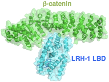
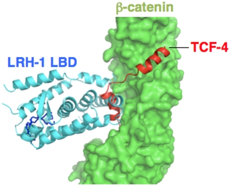
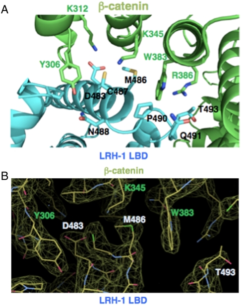
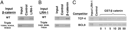
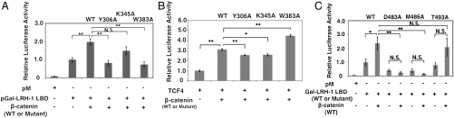
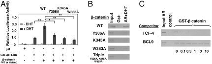
We report the three-dimensional structure of a β-catenin armadillo repeat in complex with the liver receptor homolog-1 (LRH-1) ligand binding domain at 2.8 Å resolution as the first structure of β-catenin in complex with any nuclear receptor. The surface of β-catenin that binds LRH-1 partly overlaps defined contact sites for peptide segments of β-catenin partners, including T-cell factor-4. The surface of LRH-1 that engages β-catenin is comprised of helices 1, 9, and 10 and is distinct from known interaction surfaces of LRH-1, including corepressor and coactivator binding sites. Targeted mutagenesis of amino acids forming both sides of the LRH-1/β-catenin interface reveals that they are essential for stable interactions between these proteins in solution. The LRH-1 binding site in β-catenin is also required for association with androgen receptor, providing evidence that the observed LRH-1/β-catenin interaction may be prototypic.
Conflict of interest statement
The authors declare no conflict of interest.
Figures
Similar articles
-
Structure and Dynamics of the Liver Receptor Homolog 1-PGC1α Complex.Mol Pharmacol. 2017 Jul;92(1):1-11. doi: 10.1124/mol.117.108514. Epub 2017 Mar 31. Mol Pharmacol. 2017. PMID: 28363985 Free PMC article.
-
Crystal structure of the human LRH-1 DBD-DNA complex reveals Ftz-F1 domain positioning is required for receptor activity.J Mol Biol. 2005 Dec 16;354(5):1091-102. doi: 10.1016/j.jmb.2005.10.009. Epub 2005 Oct 27. J Mol Biol. 2005. PMID: 16289203
-
Integrated Structural Modeling of Full-Length LRH-1 Reveals Inter-domain Interactions Contribute to Receptor Structure and Function.Structure. 2020 Jul 7;28(7):830-846.e9. doi: 10.1016/j.str.2020.04.020. Epub 2020 May 19. Structure. 2020. PMID: 32433991 Free PMC article.
-
Mechanistic insights from structural studies of beta-catenin and its binding partners.J Cell Sci. 2007 Oct 1;120(Pt 19):3337-44. doi: 10.1242/jcs.013771. J Cell Sci. 2007. PMID: 17881495 Review.
-
Interaction of nuclear receptors with the Wnt/beta-catenin/Tcf signaling axis: Wnt you like to know?Endocr Rev. 2005 Dec;26(7):898-915. doi: 10.1210/er.2003-0034. Epub 2005 Aug 26. Endocr Rev. 2005. PMID: 16126938 Review.
Cited by
-
HIV's Nef interacts with β-catenin of the Wnt signaling pathway in HEK293 cells.PLoS One. 2013 Oct 10;8(10):e77865. doi: 10.1371/journal.pone.0077865. eCollection 2013. PLoS One. 2013. PMID: 24130899 Free PMC article.
-
Gene expression and pathway analysis of CTNNB1 in cancer and stem cells.World J Stem Cells. 2016 Nov 26;8(11):384-395. doi: 10.4252/wjsc.v8.i11.384. World J Stem Cells. 2016. PMID: 27928465 Free PMC article.
-
Antiproliferation activity of a small molecule repressor of liver receptor homolog 1.Mol Pharmacol. 2015 Feb;87(2):296-304. doi: 10.1124/mol.114.095554. Epub 2014 Dec 3. Mol Pharmacol. 2015. PMID: 25473120 Free PMC article.
-
An animal model recapitulates human hepatic diseases associated with GATA6 mutations.Proc Natl Acad Sci U S A. 2025 Jan 7;122(1):e2317801121. doi: 10.1073/pnas.2317801121. Epub 2024 Dec 31. Proc Natl Acad Sci U S A. 2025. PMID: 39739787 Free PMC article.
-
Divergent Androgen Receptor and Beta-Catenin Signaling in Prostate Cancer Cells.PLoS One. 2015 Oct 28;10(10):e0141589. doi: 10.1371/journal.pone.0141589. eCollection 2015. PLoS One. 2015. PMID: 26509262 Free PMC article.
References
-
- Fayard E, Auwerx J, Schoonjans K. LRH-1: An orphan nuclear receptor involved in development, metabolism and steroidogenesis. Trends Cell Biol. 2004;14:250–260. - PubMed
-
- Paré JF, et al. The fetoprotein transcription factor (FTF) gene is essential to embryogenesis and cholesterol homeostasis and is regulated by a DR4 element. J Biol Chem. 2004;279:21206–21216. - PubMed
-
- Heng JC, et al. The nuclear receptor Nr5a2 can replace Oct4 in the reprogramming of murine somatic cells to pluripotent cells. Cell Stem Cell. 2010;6:167–174. - PubMed
Publication types
MeSH terms
Substances
Associated data
- Actions
Grants and funding
LinkOut - more resources
Full Text Sources
Molecular Biology Databases
