

Ceramide and mitochondria in ischemic brain injury
- PMID: 22187669
- PMCID: PMC3242427
Ceramide and mitochondria in ischemic brain injury
Abstract
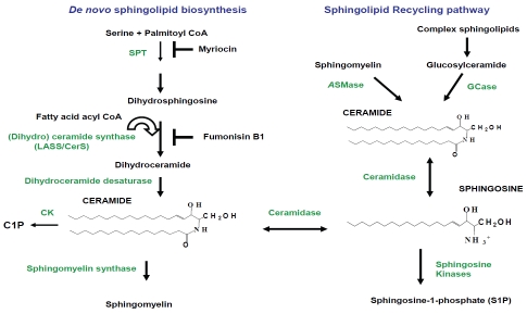
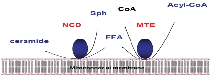
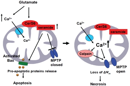
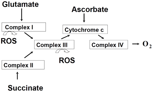
Sphingolipids are essential structural components of cellular membranes, playing prominent roles in signal transduction that governs cell proliferation, differentiation and apoptosis. Ceramides, a family of distinct molecular species characterized by various acyl chains, are synthesized de novo at the cytosolic side of the endoplasmic reticulum serving as precursors for the biosynthesis of sphingolipids in the Golgi. Recently, mitochondria emerged as an important intracellular compartment of sphingolipid metabolism. Thus, several sphingolipid-metabolizing enzymes were found to be associated with mitochondria, including neutral ceramidase, novel neutral sphingomyelinase, and (dihydro) ceramide synthase, an important ceramide-generating enzyme in de novo ceramide synthesis and recycling pathway. Mitochondrial dysfunction appears to be essential in tissue damage after brain ischemia/reperfusion (IR). Mitochondria are known to be involved in both the necrosis and apoptosis detected in animal models of ischemic stroke, and treatments that ameliorate tissue infarction were associated with better recovery of mitochondrial function. Although mitochondrial injury in stroke has been extensively studied and key mitochondrial functions affected by IR are mainly characterized, the nature of the molecule that causes loss of mitochondrial integrity and function remains obscure. Emerging data indicate a deregulation of ceramide metabolism in mitochondria damaged by IR suggesting that ceramides could play critical roles in cerebral IR-induced mitochondrial damage. This review will examine the experimental evidence supporting the key role of ceramides in mitochondrial dysfunction in cerebral IR and highlight potential targets for development of novel therapeutic approaches for stroke treatment.
Keywords: Sphingolipid; ceramide; ceramide synthase; mitochondria; neutral ceramidase; stroke.
Figures
References
-
- Hannun YA, Obeid LM. The Ceramide-centric universe of lipid-mediated cell regulation: stress encounters of the lipid kind. J Biol Chem. 2002;277:25847–50. - PubMed
-
- Ogretmen B, Hannun YA, Biologically active sphingolipids in cancer pathogenesis and treatment. Nat Rev Cancer. 2004;4:604–16. - PubMed
-
- Hannun YA, Obeid LM. Principles of bioactive lipid signalling: lessons from sphingolipids. Nat Rev Mol Cell Biol. 2008;9:139–50. - PubMed
-
- Sot J, Goni FM, Alonso A. Molecular associations and surface-active properties of short-and long-N-acyl chain ceramides. Biochim Biophys Acta. 2005;1711:12–9. - PubMed
Grants and funding
LinkOut - more resources
Full Text Sources