. 2012 Feb;18(2):230-40.
doi: 10.1261/rna.031187.111. Epub 2011 Dec 20.

Crystal structure of release factor RF3 trapped in the GTP state on a rotated conformation of the ribosome

Affiliations

Crystal structure of release factor RF3 trapped in the GTP state on a rotated conformation of the ribosome

Jie Zhou et al. RNA. 2012 Feb.

Abstract

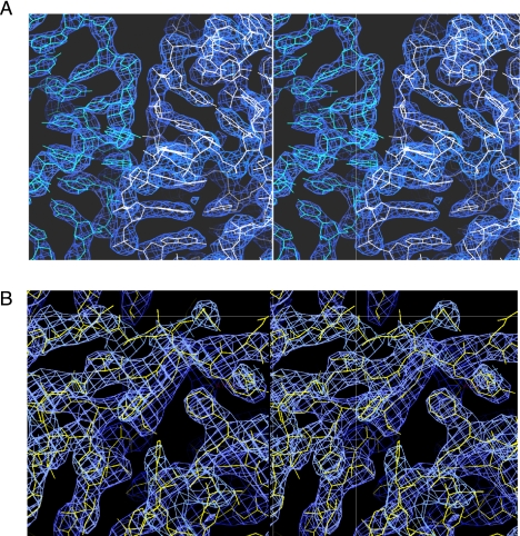
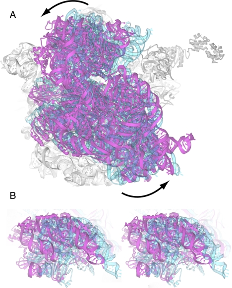
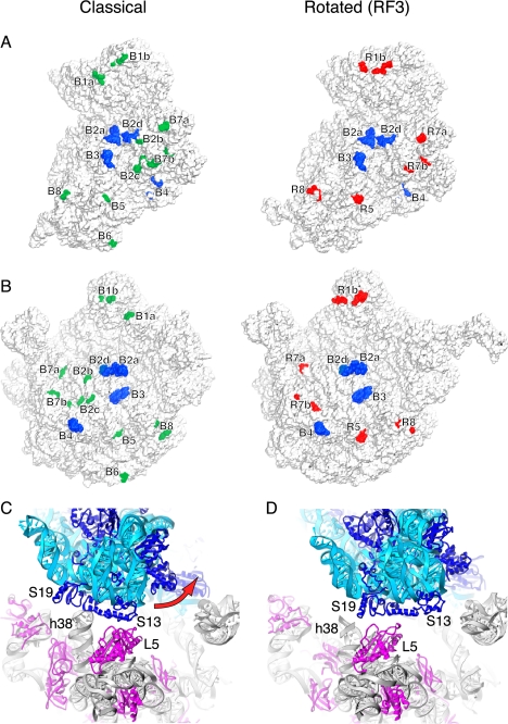
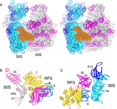
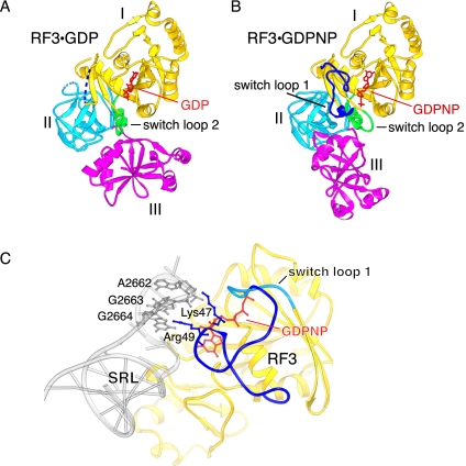
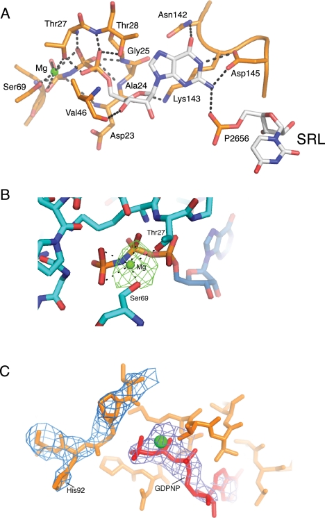
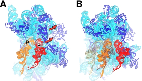
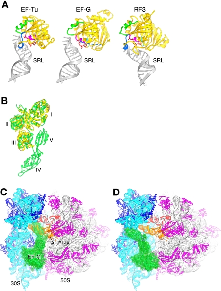
The class II release factor RF3 is a GTPase related to elongation factor EF-G, which catalyzes release of class I release factors RF1 and RF2 from the ribosome after termination of protein synthesis. The 3.3 Å crystal structure of the RF3·GDPNP·ribosome complex provides a high-resolution description of interactions and structural rearrangements that occur when binding of this translational GTPase induces large-scale rotational movements in the ribosome. RF3 induces a 7° rotation of the body and 14° rotation of the head of the 30S ribosomal subunit, and itself undergoes inter- and intradomain conformational rearrangements. We suggest that ordering of critical elements of switch loop I and the P loop, which help to form the GTPase catalytic site, are caused by interactions between the G domain of RF3 and the sarcin-ricin loop of 23S rRNA. The rotational movements in the ribosome induced by RF3, and its distinctly different binding orientation to the sarcin-ricin loop of 23S rRNA, raise interesting implications for the mechanism of action of EF-G in translocation.


Figures

FIGURE 1.
FIGURE 1.
Example of electron density maps. (A) Electron density for rRNA. Stereo view of 3.3 Å resolution 2Fo–Fc electron density contoured at 2.5 σ, showing contact between 16S rRNA (cyan, left), and 23S rRNA (gray, right), around intersubunit bridge B3. (B) Electron density map for RF3. Stereo representation of unbiased 3.3 Å resolution Fo–Fc electron density for release factor RF3, contoured at 2.4 σ.
FIGURE 2.
FIGURE 2.
Conformational changes in the ribosome in the RF3·GDPNP complex. (A) 7° counter-clockwise rotation of the 30S subunit (foreground) in the RF3 complex, as viewed from the solvent surface of the 30S subunit, relative to the classical-state RF2 complex (Korostelev et al. 2008a). The 50S subunit (gray) is in the background. (B) Stereo view showing 14° counter-clockwise rotation of the head domain of the 30S subunit, as viewed from the top of the subunit. The subunit interface is at the top of the figure. 16S rRNA and 30S proteins are colored magenta (RF3 complex) and cyan (RF2 complex).
FIGURE 3.
FIGURE 3.
Rearrangement of intersubunit bridges. (A,B) Molecular surface representations of (A) 30S subunits and (B) 50S subunits showing intersubunit bridge contacts of the classical-state RF2 70S ribosome complex (left) (Korostelev et al. 2008a) and the rotated RF3 70S complex (right). Bridge contacts are colored according to whether they are maintained (blue), disrupted (green), or newly formed (red) in the rotated state. Classical-state bridges are numbered according to Yusupov et al. (2001). (C,D) Disruption and reformation of intersubunit bridges involving the head of the 30S subunit. (C) In the classical-state RF2 complex, bridge B1a is formed between protein S13 and helix 38 of 23S rRNA, and B1b between S13 and L5. (D) In the RF3 complex, a large-scale counter-clockwise rotation of the 30S head results in disruption of bridges B1a and B1b; bridge R1b is formed between proteins S13 and S19 from the 30S subunit and L5 from the 50S subunit, involving a 34 Å displacement of the intersubunit contacts.
FIGURE 4.
FIGURE 4.
Binding of RF3 to the 70S ribosome. (A) Stereo view of RF3 (orange) bound at the entrance to the interface cavity of the 70S ribosome. (B) RF3 contacts viewed from inside the interface cavity. Domain I (the G domain; yellow) of RF3 interacts with the sarcin–ricin loop (SRL) of 23S rRNA (gray) and with protein L6 (light magenta). (C) View from inside the interface cavity, showing contacts between domains II (blue) and III (magenta) of RF3 with helix 5 of 16S rRNA (cyan) and protein S12 (dark blue).
FIGURE 5.
FIGURE 5.
Conformational rearrangements in RF3. (A,B) Comparison of crystal structures of free RF3·GDP (A) (Gao et al. 2007) and ribosome-bound RF3·GDPNP (B). A large-scale rotational movement of domain III (magenta) and smaller movements in domains I (yellow) and II (cyan) can be seen. Disordered segments in RF3·GDP are shown as dotted lines. Rearranged segments in RF3·GDPNP containing switch loops I and II are shown in blue and green, respectively. GDP and GDPNP are shown in red. (C) Ordering of residues 39–69 and repositioning of residues 70–73 of RF3 (including the switch loop 1 residues 64–72) creates a contact between the G domain of RF1 and the SRL of 23S rRNA. Residues 39–63 are shown in dark blue, the switch loop in cyan, and the rest of RF3 in yellow.
FIGURE 6.
FIGURE 6.
Interactions in the GTP binding site of RF3. (A) An intricate network of H-bonds is formed between the base, ribose, and phosphate groups of GDPNP and domain I of RF3, including elements of switch I (residues 64–72), P loop (residues 21–28), NKXD motif (residues 142–145), and phosphate 2656 of the SRL of 23S rRNA. A Mg atom (green) is coordinated by Thr27, Ser91, and the β and γ phosphates of GDPNP (see Supplemental Fig. S9). (B) Difference electron density showing Mg bound to GDPNP. An Fo–Fc difference map was calculated using the complete RF3·GDPNP·70S ribosome model, shown here contoured at 4.5 σ. The Mg ion (green) is coordinated with the β- and γ-phosphate oxygens of GDPNP and with the side-chain hydroxyls of Thr27 and Ser 69. (C) His92 of RF3 is positioned at a distance of 8 Å from the γ phosphate of GDPNP. The 2Fo–Fc electron density map is shown around His92 and GDPNP, contoured at 1.3 σ and 2.3 σ, respectively.
FIGURE 7.
FIGURE 7.
Geometry of head rotation and tRNA positioning in the 30S subunit. P-site ASL contacts in the 30S subunit head are displaced by 23 Å in the RF3 complex, relative to their position in the classical-state RF2 complex. Docking of P-site (orange) and E-site (red) tRNAs on the (A) classical-state RF2 and (B) rotated RF3 structures illustrates that the combined 30S head and body rotations seen in the RF3 structure are sufficient to translocate an ASL from the 30S P site to the E site. The views in A and B are shown in exactly the same orientation relative to the 50S subunit. In B, the initial positions of the P- and E-site tRNAs as in A are shown in transparent orange and red, respectively.
FIGURE 8.
FIGURE 8.
Implications of the orientation of RF3 domain I for EF-G binding. (A) Orientations of domain I of EF-Tu (Voorhees et al. 2010), EF-G (Gao et al. 2009), and RF3 (this work) relative to the SRL of 23S rRNA. The domains were aligned in three dimensions by virtue of their conserved structural elements. The conserved functional elements of the respective factors are indicated: switch 1 (blue); switch 2 (green); P loop (magenta); NKXD motif (cyan). The G nucleotides are shown in red. The orientation of the SRL in in the RF3 complex is rotated by ∼45° relative to those of the other factors. (B) Three-dimensional alignment of Domains I, II, and III of RF3·GDPNP (yellow) and EF-G (green) (Gao et al. 2009) show clear structural homology. (C) Observed orientation of EF-G (green) bound to the classical-state 70S ribosome·EF-G complex (Gao et al. 2009), in which an A-site tRNA has been docked in the A site; note the steric clash with A-site tRNA. (D) Orientation of EF-G in the classical-state ribosome with its domain I docked on the 50S subunit in the same orientation as that of RF3·GDPNP. Note the absence of steric clash with A-site tRNA or the ribosome.

Comment in

Similar articles

Cited by

References

    1. Adams PD, Afonine PV, Bunkóczi G, Chen VB, Davis IW, Echols N, Headd JJ, Hung LW, Kapral GJ, Grosse-Kunstleve RW, et al. 2010. PHENIX: A comprehensive Python-based system for macromolecular structure solution. Acta Crystallogr D Biol Crystallogr 66: 213–221 - PMC - PubMed
    1. Agirrezabala X, Lei J, Brunelle JL, Ortiz-Meoz RF, Green R, Frank J 2008. Visualization of the hybrid state of tRNA binding promoted by spontaneous ratcheting of the ribosome. Mol Cell 32: 190–197 - PMC - PubMed
    1. Allen GS, Zavialov A, Gursky R, Ehrenberg M, Frank J 2005. The cryo-EM structure of a translation initiation complex from Escherichia coli. Cell 121: 703–712 - PubMed
    1. Ben-Shem A, Jenner L, Yusupova G, Yusupov M 2010. Crystal structure of the eukaryotic ribosome. Science 330: 1203–1209 - PubMed
    1. Berk V, Zhang W, Pai RD, Cate JH 2006. Structural basis for mRNA and tRNA positioning on the ribosome. Proc Natl Acad Sci 103: 15830–15834 - PMC - PubMed

Publication types

MeSH terms

LinkOut - more resources