Review
. 2012 Jan 15;441(2):523-40.
doi: 10.1042/BJ20111451.

Autophagy, mitochondria and oxidative stress: cross-talk and redox signalling

Affiliations
Review

Autophagy, mitochondria and oxidative stress: cross-talk and redox signalling

Jisun Lee et al. Biochem J. .

Abstract

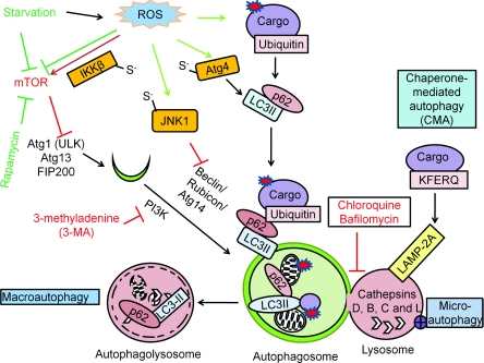
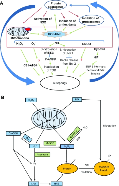
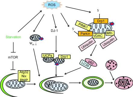
Reactive oxygen and nitrogen species change cellular responses through diverse mechanisms that are now being defined. At low levels, they are signalling molecules, and at high levels, they damage organelles, particularly the mitochondria. Oxidative damage and the associated mitochondrial dysfunction may result in energy depletion, accumulation of cytotoxic mediators and cell death. Understanding the interface between stress adaptation and cell death then is important for understanding redox biology and disease pathogenesis. Recent studies have found that one major sensor of redox signalling at this switch in cellular responses is autophagy. Autophagic activities are mediated by a complex molecular machinery including more than 30 Atg (AuTophaGy-related) proteins and 50 lysosomal hydrolases. Autophagosomes form membrane structures, sequester damaged, oxidized or dysfunctional intracellular components and organelles, and direct them to the lysosomes for degradation. This autophagic process is the sole known mechanism for mitochondrial turnover. It has been speculated that dysfunction of autophagy may result in abnormal mitochondrial function and oxidative or nitrative stress. Emerging investigations have provided new understanding of how autophagy of mitochondria (also known as mitophagy) is controlled, and the impact of autophagic dysfunction on cellular oxidative stress. The present review highlights recent studies on redox signalling in the regulation of autophagy, in the context of the basic mechanisms of mitophagy. Furthermore, we discuss the impact of autophagy on mitochondrial function and accumulation of reactive species. This is particularly relevant to degenerative diseases in which oxidative stress occurs over time, and dysfunction in both the mitochondrial and autophagic pathways play a role.


Figures

Figure 1
Figure 1. Autophagy mechanisms and regulation
There are three different mechanisms of autophagy: (i) macroautophagy, the bulk degradation of cargo; (ii) microautophagy, involution of the lysosome around cargo; and (iii) chaperone-mediated autophagy (CMA), the degradation of the tagged cargo. The autophagosome and the lysosome fuse to degrade the cargo by lysosomal acidic hydrolases, including cathepsins D, B and L. Macroautophagy is regulated by starvation through the Atg1–Atg13 complex, which is inhibited by mTOR activation. Rapamycin or nutrient starvation causes mTOR inactivation, subsequent Atg1–Atg13 activation and activates macroautophagy. The beclin-1–PI3K complex is involved in autophagosomal expansion. 3-MA inhibits PI3K activity. Atg8–pro-LC3 is cleaved by Atg4, modified by PE to become LC3-II and inserted into the autophagosomes. The puncta formed by LC3-II and the electrophoretic separation of LC3-I and LC3-II are used as a marker for autophagosomal accumulation. RFP–GFP–LC3 (tfLC3) form puncta with both red and green fluorescence in the autophagosomes, and only red puncta when the autophagosomes fuse with lysosomes where GFP is inactivated, therefore tfLC3 is used to monitor autophagic flux. The degradation rate of long-lived proteins is another method for measurement of autophagic flux. p62 binds both LC3 and ubiquitin and therefore plays a role in targeting ubiquitinated proteins to the autophagosomes. Red arrows are indicative of negative regulators of autophagy, and green arrows are indicative of positive regulators of autophagy. Atg4 Cys81 thiol modification is involved in starvation and H2O2-induced autophagy. S-nitrosation of IKKβ and JNK1 are involved in NO-induced inhibition of autophagy.
Figure 2
Figure 2. ROS/RNS production and signalling
(A) ROS/RNS production and signalling of autophagy. ROS and RNS production can be induced by protein aggregates, generated by the mitochondrial respiratory activities and by other cellular oxidases. These ROS/RNS species can modify signalling molecules to stimulate or inhibit autophagy. Atg4 Cys81 thiol modification is involved in starvation and H2O2-induced autophagy. S-nitrosation of IKKβ and JNK1 are involved in NO-induced inhibition of autophagy. Although many ROS/RNS have effects on autophagy, besides a handful of known ROS/RNS-targeted autophagy regulators, the exact mechanisms for the effect of ROS/RNS on autophagy are largely unknown. (B) Mitochondrial production of ROS/RNS. Regardless of their sources, H2O2 and NO can target to the mitochondria. Mitochondrially produced superoxide (O2•−) can react with NO to produce peroxynitrite (ONOOH). O2•−, H2O2 and ONOOH can further damage the mitochondria in combination with iron and produce lipid peroxidation (LPO) products, including HNE. MnSOD, aconitase, GPX (glutathione peroxidase) and catalase are involved in generation and reduction of these products. HNE, NO and H2O2 can modify proteins to initiate signalling mechanisms or damage. The most sensitive moieties to oxidative modification are the thiols on cysteine residues. Redox reactions in protein modification are involved in signalling of autophagy.
Figure 3
Figure 3. Mitophagy
Mitophagy is the specific degradation of the mitochondria in response to global signals, including starvation and oxidative stress, or specific signals including mitochondrial targeting of signalling proteins or modification of mitochondrial proteins. In yeast, Atg32 is specific for mitophagy by targeting the mitochondria to the autophagosome. In mammalian cells, Nix is involved in mitochondrial clearance during erythrocyte maturation. In response to reduced cellular ATP, AMPK is activated and phosphorylates ULK1 and ULK2 (two Atg1 homologues) to activate both general macroautophagy and mitophagy. Parkinson's disease genes encoding α-synuclein, parkin, PINK1 and DJ-1 are all involved in mitophagy. A decrease in mitochondrial membrane potential (ψm) can be induced by ROS, and by targeting α-synuclein to the mitochondria. This decrease in mitochondrial membrane potential serves as a signal for mitophagy. In addition to a decrease in mitochondrial membrane potential, mitochondrial fission is another signal for mitophagy. PINK1 facilitates parkin targeting to the mitochondria, and ubiquitinates the mitochondrial outer membrane protein VDAC. Ubiquitinated VDAC can be recognized by p62 to initiate mitophagy. DJ-1 senses oxidative stress and serves a parallel pathway to maintain mitochondrial membrane potential and preserve mitochondria from fragmentation. Many of the regulators of mitophagy can be regulated by ROS. For example, α-synuclein is nitrated and, as a consequence, increases its aggregation propensity. Parkin can be sulfonated and S-nitrosated. Drp1 S-nitrosation is also involved in regulation of mitochondrial fission and associated induction of mitophagy.

References

    1. de Duve C. The lysosome. Sci. Am. 1963;208:64–72. - PubMed
    1. de Duve C., Wattiaux R. Functions of lysosomes. Annu. Rev. Physiol. 1966;28:435–492. - PubMed
    1. Shacka J. J., Roth K. A., Zhang J. The autophagy–lysosomal degradation pathway: role in neurodegenerative disease and therapy. Front. Biosci. 2008;13:718–736. - PubMed
    1. Schapira A. H., Gegg M. Mitochondrial contribution to Parkinson's disease pathogenesis. Parkinsons Dis. 2011;2011:159160. - PMC - PubMed
    1. Crifo C., Capuozzo E., Siems W., Salerno C. Inhibition of ion transport ATPases by HNE. Biofactors. 2005;24:137–140. - PubMed

Publication types

MeSH terms