. 2011 Dec 21:4:44.
doi: 10.1186/1756-6606-4-44.

Dependence of NMDA/GSK-3β mediated metaplasticity on TRPM2 channels at hippocampal CA3-CA1 synapses

Affiliations

Dependence of NMDA/GSK-3β mediated metaplasticity on TRPM2 channels at hippocampal CA3-CA1 synapses

Yu-Feng Xie et al. Mol Brain. .

Abstract

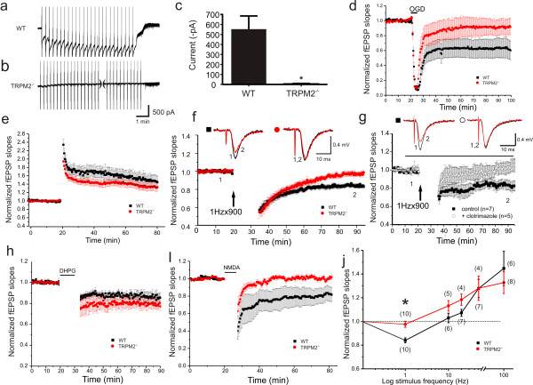
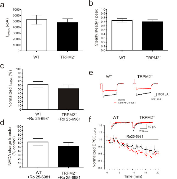
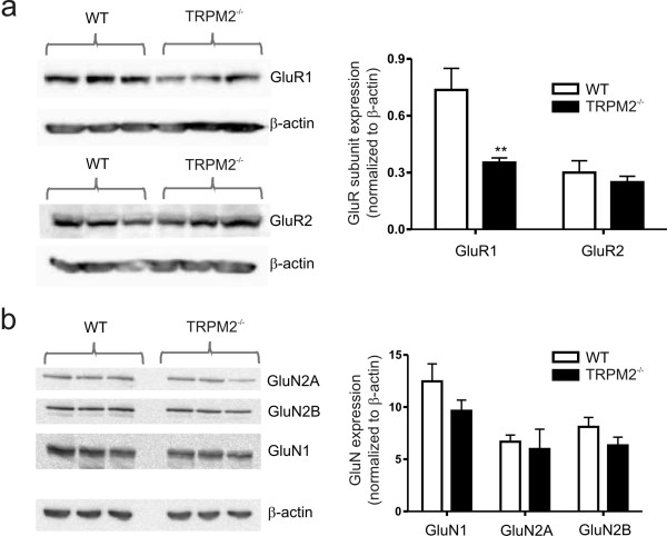
Transient receptor potential melastatin 2 (TRPM2) is a calcium permeable non-selective cation channel that functions as a sensor of cellular redox status. Highly expressed within the CNS, we have previously demonstrated the functional expression of these channels in CA1 pyramidal neurons of the hippocampus. Although implicated in oxidative stress-induced neuronal cell death, and potentially in neurodegenerative disease, the physiological role of TRPM2 in the central nervous system is unknown. Interestingly, we have shown that the activation of these channels may be sensitized by co-incident NMDA receptor activation, suggesting a potential contribution of TRPM2 to synaptic transmission. Using hippocampal cultures and slices from TRPM2 null mice we demonstrate that the loss of these channels selectively impairs NMDAR-dependent long-term depression (LTD) while sparing long-term potentiation. Impaired LTD resulted from an inhibition of GSK-3β, through increased phosphorylation, and a reduction in the expression of PSD95 and AMPARs. Notably, LTD could be rescued in TRPM2 null mice by recruitment of GSK-3β signaling following dopamine D2 receptor stimulation. We propose that TRPM2 channels play a key role in hippocampal synaptic plasticity.


Figures

Figure 1
Figure 1
LTD is impaired in hippocampal slices derived from TRPM2-/- mice. (a,b) ADPR-primed TRPM2 currents are facilitated by voltage ramps ( ± 100 mV, 1/10 sec) in WT (a), but not in cultured neurons from TRPM2-/- mice (b). (c) Summary bar graph of ADPR-primed peak current amplitude in WT and TRPM2-/- neurons. (unpaired t-test p = 0.001, 553.4 ± 132 pA in WT, n = 5; 7.9 ± 7.2 pA in KO, n = 6) (d) Transient oxygen-glucose deprivation (5 min OGD) causes long-lasting depression of fEPSP slopes in slices from WT (n = 12) but not TRPM2-/- (n = 6). (e) LTP is unaffected by knockout of TRPM2 (WT, n = 6; TRPM2-/-, n = 8). (f) LTD of fEPSPs evoked by repetitive stimulation (900 stimuli at 1 Hz) in WT slices (n = 10) is absent in slices from TRPM2-/- mice (n = 10). (g) LTD of fEPSPs was inhibited by application of clotrimazole, a TRPM2 inhibitor. Timing of clotrimazole application, in treated slices, is indicated by the black bar. (h) Metabotropic-glutamate receptor dependent LTD is unimpaired by deletion of TRPM2 (WT, n = 6; TRPM2-/-, n = 7). (i) Chem-LTD, evoked by 5 min application of NMDA (10 μM) is abolished in slices from TRPM2-/- (n = 6) but not WT (n = 6) slices. (j) Summary graph for a series of recordings from WT and TRPM2-/- slices in which plasticity was induced by repetitive stimulation delivered at 1, 10, 20, 50 Hz (900 pulses at each frequency) or 100 Hz (four 1 sec trains delivered 20 sec apart). For each induction frequency, normalized fEPSP slopes, measured at the end of recording from each of the two populations of slices, are plotted. Numbers in parentheses represents the number of recordings at each point. *denotes p = 0.016.
Figure 2
Figure 2
NMDAR function is unaffected by disruption of TRPM2 channel expression. Neither peak (a) nor steady-state/peak (b) currents, generated in response to rapid applications of NMDA to cultured hippocampal neurons, are affected by loss of TRPM2 expression. Similarly, the sensitivity of NMDA-evoked responses (peak amplitude (c), total charge transfer (d) and representative traces (e) recorded from cultured hippocampal neurons, to inhibition by the GluN2B specific antagonist, Ro 25-6981, is unaltered in neurons from TRPM2-/-. (f) Inhibition of pharmacologically isolated NMDAR-mediated synaptic currents, recorded from CA1 pyramidal neurons in response to Schaffer-collateral stimulation, by Ro 25-6981 is unaltered in slices from TRPM2-/-. For all results presented in panels a-d, n = 5; for panel f, WT: n = 6; TRPM2-/-: n = 5)
Figure 3
Figure 3
Inhibition of GSK-3β phosphorylation and reduced PSD95 and AMPAR expression contributes to impaired LTD in TRPM2-/- mice. (a) The ratio of AMPAR- to NMDAR-mediated EPSCs is reduced in TRPM2-/- neurons. (b,c) The amplitude, but not the frequency, of mEPSCs is reduced in slices from TRPM2-/- mice. Representative traces of mEPSCs recordings from slices derived from WT and TRPM2-/- mice are shown. The expression of (d) PSD-95 (normalized to β-actin loading control) and (e) of the AMPAR subunit, GluR1, is depressed in slices from TRPM2-/- mice. (f) Knockout of TRPM2 causes increased phosphorylation of GSK-3β, at its inhibitory site (Ser9, n = 3). (g) Insulin application to hippocampal slices depresses AMPAR-mediated fEPSPs in slices from TRPM2-/-, but not WT, slices. (h) Simulation of dopamine D2 receptors by quinpirole (10 μM) reduces GSK-3β phosphorylation at Ser9 (n = 3). (i) D2 receptor stimulation by quinpirole rescues LTD in slices from TRPM2-/- mice.
Figure 4
Figure 4
Comparison of glutamate receptor subunit expression in hippocampal slices derived from WT and TRPM2-/- mice. (a) Western blots and summary bar graph representing the expression levels of the AMPA receptor subunit subtypes, GluR1 and GluR2. Note that the quantified data for GluR1 is the same as that presented in Figure 2. (b) Western blots and summary bar graph representing the expression levels of the NMDA receptor subunit subtypes, GluN1 (n = 3), GluN2A (n = 5) and GluN2B (n = 3).

References

    1. Takahashi N, Kozai D, Kobayashi R, Ebert M, Mori Y. Roles of TRPM2 in oxidative stress. Cell Calcium. 2011. - PubMed
    1. Xie YF, Macdonald JF, Jackson MF. TRPM2, calcium and neurodegenerative diseases. Int J Physiol Pathophysiol Pharmacol. 2010;2(2):95–103. - PMC - PubMed
    1. Olah ME, Jackson MF, Li H, Perez Y, Sun HS, Kiyonaka S, Mori Y, Tymianski M, MacDonald JF. Ca2+-dependent induction of TRPM2 currents in hippocampal neurons. J Physiol. 2009;587(Pt 5):965–979. - PMC - PubMed
    1. Scharenberg AM. TRPM2 and TRPM7: channel/enzyme fusions to generate novel intracellular sensors. Pflugers Arch. 2005;451(1):220–227. doi: 10.1007/s00424-005-1444-0. - DOI - PubMed
    1. Yamamoto S, Shimizu S, Kiyonaka S, Takahashi N, Wajima T, Hara Y, Negoro T, Hiroi T, Kiuchi Y, Okada T, Kaneko S, Lange I, Fleig A, Penner R, Nishi M, Takeshima H, Mori Y. TRPM2-mediated Ca2+influx induces chemokine production in monocytes that aggravates inflammatory neutrophil infiltration. Nat Med. 2008;14(7):738–747. doi: 10.1038/nm1758. - DOI - PMC - PubMed

Publication types

MeSH terms