Review
. 2012 Feb;23(1):83-8.
doi: 10.1016/j.copbio.2011.11.023. Epub 2011 Dec 19.

Single-cell protein analysis

Affiliations
Review

Single-cell protein analysis

Meiye Wu et al. Curr Opin Biotechnol. 2012 Feb.

Abstract

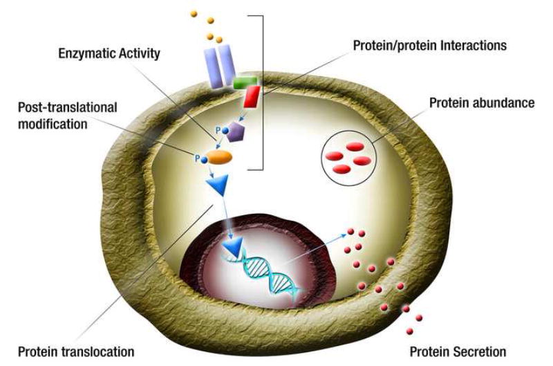
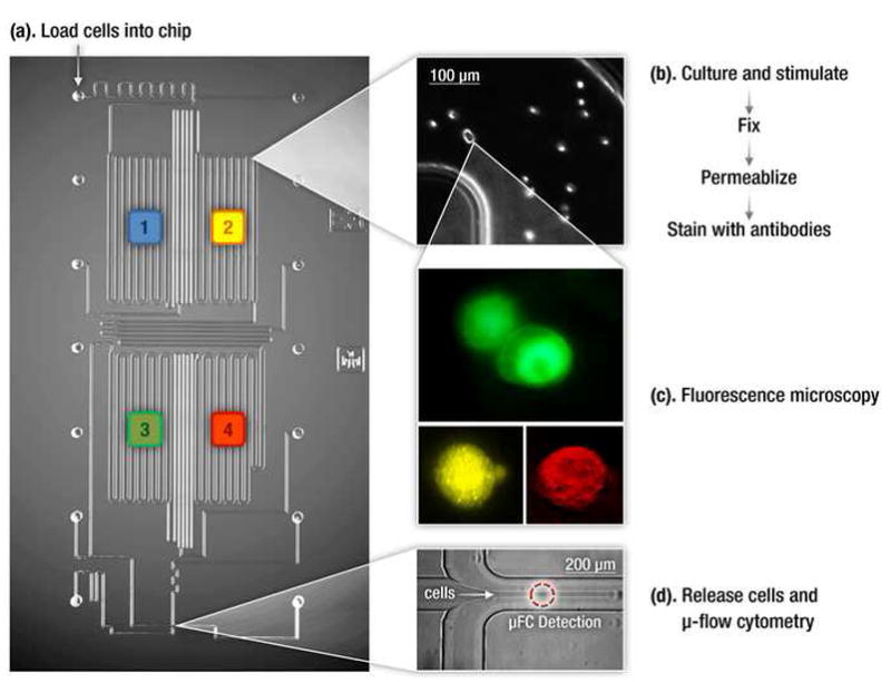
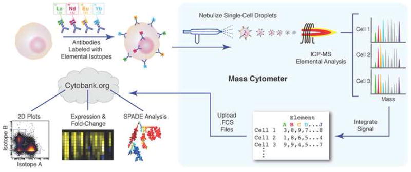
Heterogeneity of cellular systems has been widely recognized but only recently have tools become available that allow probing of genes and proteins in single cells to understand it. While the advancement in single cell genomic analysis has been greatly aided by the power of amplification techniques (e.g. PCR), analysis of proteins in single cells has proven to be more challenging. However, recent advances in multi-parameter flow cytometry, microscopy, microfluidics and other techniques have made it possible to measure wide variety of proteins in single cells. In this review, we highlight key recent developments in analysis of proteins in a single cell (excluding imaging-based methods), and discuss their significance in biological research.


Figures

Figure 1
Figure 1. Protein analysis in single cells
Proteome of a cell is complex and requires measurements of many attributes including abundance or concentration of cytosolic, membrane-bound and secreted proteins, protein interactions with other proteins and molecules, inter- and intra-organelle translocation of proteins, post-translational modifications, and enzymatic and other activities of a protein. Colored shapes represent different proteins. P – phosphorylation.
Figure 2
Figure 2. Microfluidic Platform for single cell protein analysis
An integrated microfluidic platform that integrates cell culture and sample preparation with two orthogonal single-cell resolution techniques- flow cytomtery and fluorescence microscopy. [18].
Figure 3
Figure 3. Mass cytometry measurement of immune cell response
Cells are stained with epitope-specific antibodies conjugated to transition element isotope reporters of different masses. Cells are nebulized into single-cell droplets, and an elemental mass spectrum is acquired for each. The integrated integrated elemental reporter signals can then be interpreted using conventional flow cytometry data analysis methods and plotted as heat maps or tree plots. (Reproduced with permission from Bendall et. al. [21])

References

    1. Spiller DG, Wood CD, Rand DA, White MR. Measurement of single-cell dynamics. Nature. 2010;465:736–745. - PubMed
    1. Spencer SL, Gaudet S, Albeck JG, Burke JM, Sorger PK. Non-genetic origins of cell-to-cell variability in TRAIL-induced apoptosis. Nature. 2009;459:428–432. - PMC - PubMed
    1. Tay S, Hughey JJ, Lee TK, Lipniacki T, Quake SR, Covert MW. Single-cell NF-kappaB dynamics reveal digital activation and analogue information processing. Nature. 2010;466:267–271. - PMC - PubMed
    1. Batchelor E, Loewer A, Lahav G. The ups and downs of p53: understanding protein dynamics in single cells. Nat Rev Cancer. 2009;9:371–377. - PMC - PubMed
    1. Taylor RJ, Falconnet D, Niemisto A, Ramsey SA, Prinz S, Shmulevich I, Galitski T, Hansen CL. Dynamic analysis of MAPK signaling using a high-throughput microfluidic single-cell imaging platform. Proc Natl Acad Sci U S A. 2009;106:3758–3763. - PMC - PubMed

Publication types