. 2012 Feb;180(2):484-93.
doi: 10.1016/j.ajpath.2011.10.005. Epub 2011 Dec 20.

Participation of miR-200 in pulmonary fibrosis

Affiliations

Participation of miR-200 in pulmonary fibrosis

Shanzhong Yang et al. Am J Pathol. 2012 Feb.

Abstract

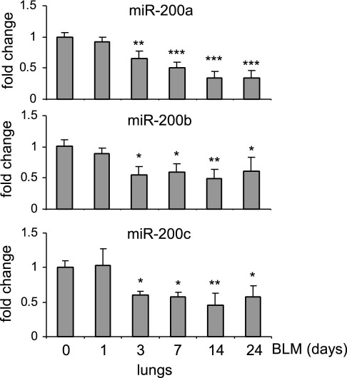
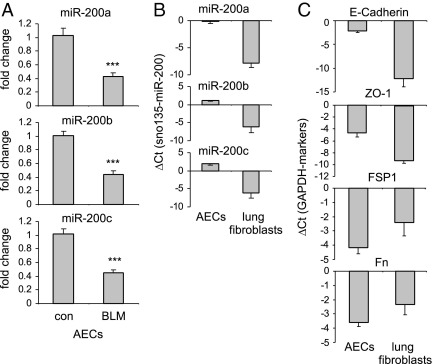
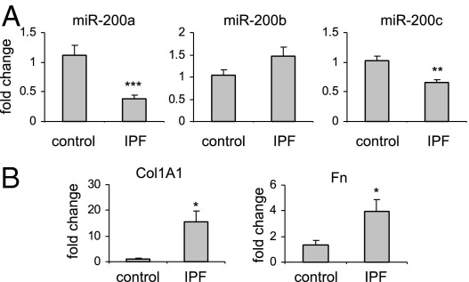
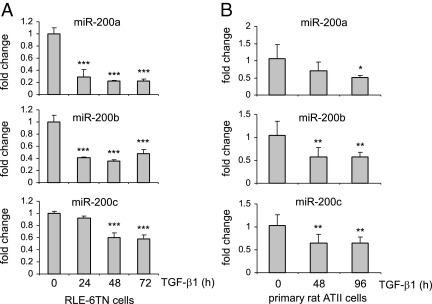
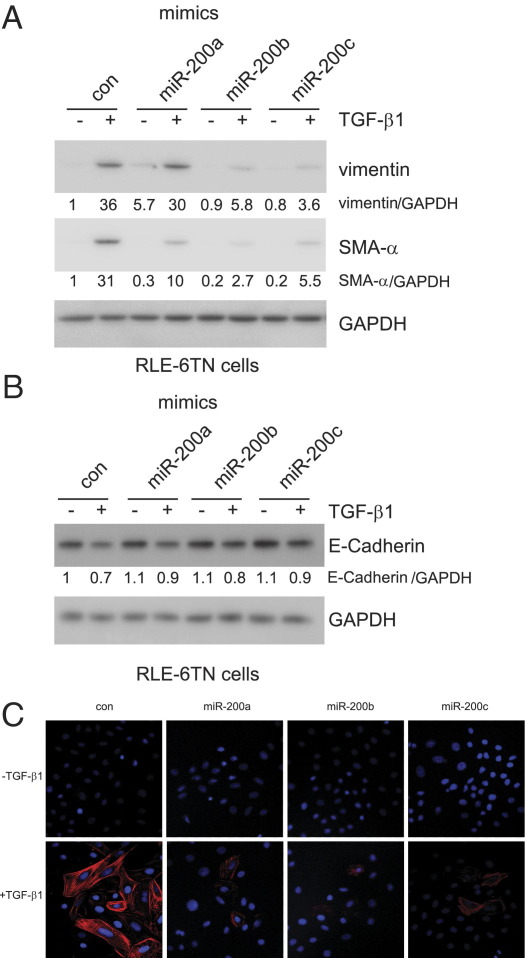
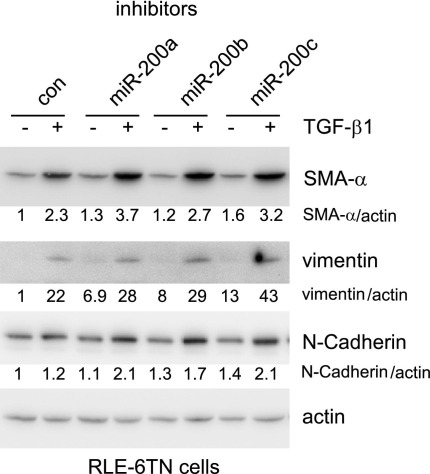
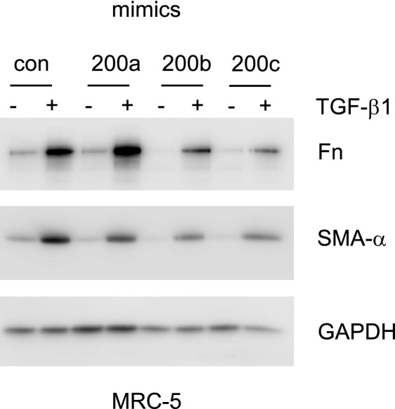
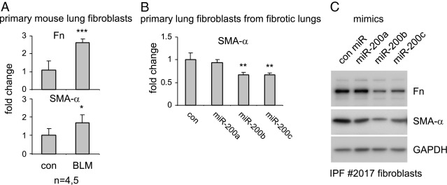
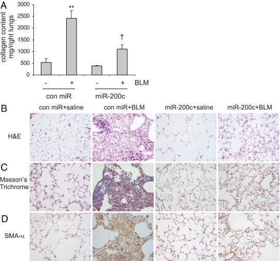
Excessive extracellular matrix production by fibroblasts in response to tissue injury contributes to fibrotic diseases, such as idiopathic pulmonary fibrosis (IPF). Epithelial-mesenchymal transition, involving transition of alveolar epithelial cells (AECs) to pulmonary fibroblasts, appears to be an important contributory process to lung fibrosis. Although aberrant expression of microRNAs (miRs) is involved in a variety of pathophysiologic processes, the role of miRs in fibrotic lung diseases is less well understood. In the present study, we found that miR-200a, miR-200b, and miR-200c are significantly down-regulated in the lungs of mice with experimental lung fibrosis. Levels of miR-200a and miR-200c were reduced in the lungs of patients with IPF. miR-200 had greater expression in AECs than in lung fibroblasts, and AECs from mice with experimental pulmonary fibrosis had diminished expression of miR-200. We found that the miR-200 family members inhibit transforming growth factor-β1-induced epithelial-mesenchymal transition of AECs. miR-200 family members can reverse the fibrogenic activity of pulmonary fibroblasts from mice with experimental pulmonary fibrosis and from patients with IPF. Indeed, the introduction of miR-200c diminishes experimental pulmonary fibrosis in mice. Thus, the miR-200 family members participate importantly in fibrotic lung diseases and suggest that restoring miR-200 expression in the lungs may represent a novel therapeutic approach in treating pulmonary fibrotic diseases.


Figures

Figure 1
Figure 1
miR-200 is down-regulated in the lungs of mice with experimental pulmonary fibrosis. Mice were injected intratracheally with saline or bleomycin (BLM; 1 U/kg in 50 μL of saline) and sacrificed at days 0, 1, 3, 4, 7, 14, or 24 after injection. Total RNA from the lungs was isolated, and the levels of miR-200a, miR-200b, and miR-200c were determined by real-time PCR assays. A small nucleolar RNA, snoRNA 135, was used as an internal control (n = 3 mice in each group). Data are given as mean ± SD. *P < 0.05, **P < 0.01, and ***P < 0.001 versus day 0.
Figure 2
Figure 2
miR-200 demonstrates greater expression in AECs than in lung fibroblasts and is down-regulated in AECs isolated from fibrotic mouse lungs. Mice were injected intratracheally with saline or bleomycin (BLM; 1 U/kg in 50 μL of saline) and sacrificed 14 days after the injection. AECs and pulmonary fibroblasts were isolated and cultured as described in Materials and Methods, and RNA from these cells was isolated. A: The levels of miR-200a, miR-200b, and miR-200c in the cells were determined by real-time PCR. con indicates control. B: The levels of miR-200a, miR-200b, and miR-200c in the AECs and pulmonary fibroblasts were determined by real-time PCR and are shown as ΔCT relative to those of a small nucleolar RNA, snoRNA 135, in the same cells. C: The levels of E-cadherin, ZO-1, FSP1, and Fn in the AECs and pulmonary fibroblasts were determined by real-time PCR and are shown as ΔCTrelative to those of GAPDH in the same cells (n = 5 mice per group). Data are given as mean ± SD. ***P < 0.001 versus the con group.
Figure 3
Figure 3
miR-200 is down-regulated in the lungs of patients with IPF. A: Total RNA was isolated from histologically normal human lungs and from the lungs of patients with IPF. The levels of miR-200a, miR-200b, and miR-200c in the lungs were determined by real-time PCR. A small nucleolar RNA, RNU48, was used as an internal control (n = 8 for healthy controls, and n = 18 for IPF lungs). Data are given as mean ± SEM. B: The expression of collagen 1A1 (Col1A1) and Fn was determined in the same RNA samples. *P < 0.05, **P < 0.01, and ***P < 0.001 versus the control group.
Figure 4
Figure 4
miR-200 is down-regulated in TGF-β1–treated AECs. A: RLE-6TN cells were starved in media containing 0.5% FBS for 24 hours and then treated with 10 ng/mL TGF-β1 for 0, 24, 48, or 72 hours. RNA was isolated, and the levels of miR-200a, miR-200b, and miR-200c in the cells were determined by real-time PCR. Experiments were performed in triplicate. Data are given as mean ± SD. ***P < 0.001 versus time 0. B: Rat primary ATII cells were isolated and cultured as described in Materials and Methods. The cells were treated with 10 ng/mL TGF-β1 for 0, 48, or 96 hours. RNA was isolated, and the levels of miR-200a, miR-200b, and miR-200c in the cells were determined by real-time PCR. Each condition contained six replicates. Data are given as mean ± SD. *P < 0.05, **P < 0.01 versus time 0.
Figure 5
Figure 5
Overexpression of miR-200 diminishes EMT in AECs. A and B: RLE-6TN cells were transfected with 40 nmol/L control (con) miR mimics or mimics for miR-200a, miR-200b, or miR-200c. Three days after the transfection, the cells were starved in medium containing 0.5% FBS for 24 hours and then treated with 5 ng/mL TGF-β1 for 48 hours. The cells were collected, and cell extracts were prepared. The levels of vimentin, SMA-α, E-cadherin, and the loading con, GAPDH, were determined by using Western blot analysis. C: RLE-6TN cells, plated on coverslips, were transfected and treated as in A and B. The cells were then fixed and permeabilized. Immunofluorescence assays were performed with anti-SMA-α antibody. Nuclei were stained with DAPI. Original magnification, ×40.
Figure 6
Figure 6
Inhibition of miR-200 enhances EMT in AECs. RLE-6TN cells were transfected with 40 nmol/L control (con) miR inhibitors or inhibitors to miR-200a, miR-200b, or miR-200c. Three days after the transfection, the cells were starved in media containing 0.5% FBS for 24 hours and then treated with 5 ng/mL TGF-β1 for 48 hours. The cells were collected, and cell extracts were prepared. The levels of vimentin, SMA-α, N-cadherin, and the loading control, actin, were determined by using Western blot analysis.
Figure 7
Figure 7
miR-200 diminishes the fibrogenic activity of TGF-β1 in lung fibroblasts. Primary human lung fibroblasts were transfected with 40 nmol/L control (con) miR mimics or mimics for miR-200a, miR-200b, or miR-200c. Three days after transfection, the cells were starved in medium containing 0.1% FBS for 24 hours and then treated with 2 ng/mL TGF-β1 for 48 hours. The cells were collected, and cell extracts were prepared. The levels of Fn, SMA-α, and the loading control, GAPDH, were determined by using Western blot analysis.
Figure 8
Figure 8
miR-200 attenuates the fibrogenic activity of lung fibroblasts from mice with experimental pulmonary fibrosis and from a patient with IPF. A: Primary lung fibroblasts from mice that were intratracheally injected with saline or bleomycin (BLM) were isolated and, after culture, RNA from these cells was purified. The expression of Fn and SMA-α was determined by real-time PCR assays. GAPDH was used as an internal control (con). *P < 0.05, ***P < 0.001 versus the con group. B: One fibroblast line from a mouse with experimental pulmonary fibrosis was transfected with 40 nmol/L con miR mimics or mimics for miR-200a, miR-200b, or miR-200c. Three days after the transfection, the cells were collected and RNA was isolated. The expression of SMA-α was determined by real-time PCR. Experiments were repeated with a second fibroblast line from fibrotic mouse lungs and showed the same results. **P < 0.001 versus the con group. C: IPF lung fibroblasts were transfected with 40 nmol/L con miR mimics or mimics for miR-200a, miR-200b, or miR-200c. Three days after the transfection, the cells were collected and cell extracts were prepared. The levels of Fn, SMA-α, and the loading con, GAPDH, were determined by using Western blot analysis.
Figure 9
Figure 9
Introduction of miR-200 into lungs diminishes bleomycin (BLM)–induced pulmonary fibrosis. A: Control (con) mimics, con mimics plus bleomycin, miR-200c mimics, or miR-200c mimics plus BLM were instilled intratracheally through oropharyngeal cavities. At 14 days after the treatment, collagen contents in the right lungs were determined by Sircol assays (n = 3, 9, 3, and 9, respectively). Data are given as mean±SEM. **P < 0.01 versus con miR-; P < 0.01 versus con miR+. BD: The experiments were performed as in A. Lung tissue sections were prepared, and H&E staining (B), Masson's trichrome staining (C), and IHC staining for SMA-α (D) were performed. Original magnification, ×40.

Similar articles

Cited by

References

    1. Tomasek J.J., Gabbiani G., Hinz B., Chaponnier C., Brown R.A. Myofibroblasts and mechano-regulation of connective tissue remodelling. Nat Rev Mol Cell Biol. 2002;3:349–363. - PubMed
    1. Wynn T.A. Common and unique mechanisms regulate fibrosis in various fibroproliferative diseases. J Clin Invest. 2007;117:524–529. - PMC - PubMed
    1. Thannickal V.J., Toews G.B., White E.S., Lynch J.P., 3rd, Martinez F.J. Mechanisms of pulmonary fibrosis. Annu Rev Med. 2004;55:395–417. - PubMed
    1. Hardie W.D., Glasser S.W., Hagood J.S. Emerging concepts in the pathogenesis of lung fibrosis. Am J Pathol. 2009;175:3–16. - PMC - PubMed
    1. Chapman H.A. Epithelial-mesenchymal interactions in pulmonary fibrosis. Annu Rev Physiol. 2011;73:413–435. - PubMed

Publication types

MeSH terms