. 2012 Apr;40(8):3456-69.
doi: 10.1093/nar/gkr1242. Epub 2011 Dec 20.

Cdx2 homeoprotein inhibits non-homologous end joining in colon cancer but not in leukemia cells

Affiliations

Cdx2 homeoprotein inhibits non-homologous end joining in colon cancer but not in leukemia cells

Benjamin Renouf et al. Nucleic Acids Res. 2012 Apr.

Abstract

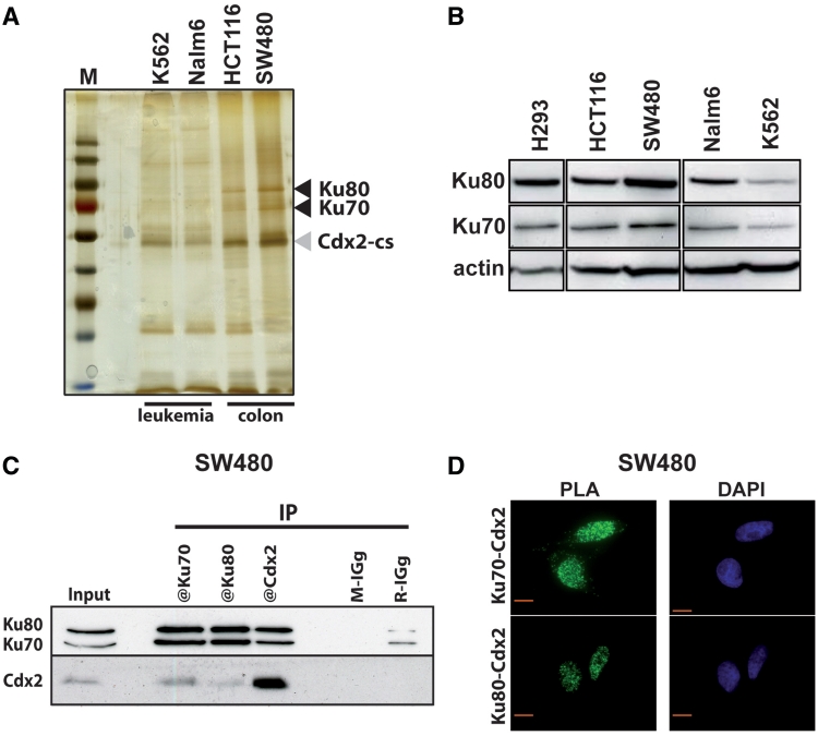
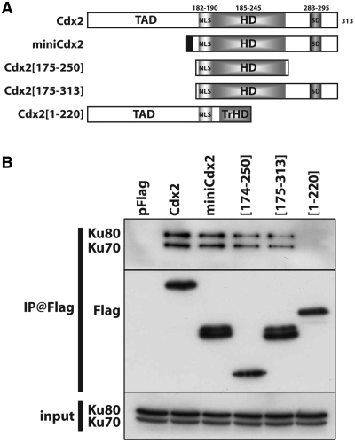
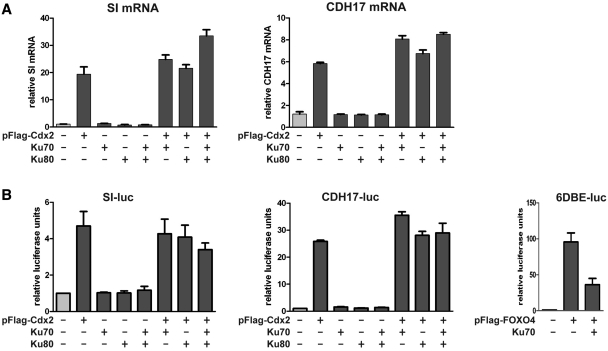
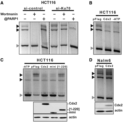
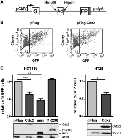
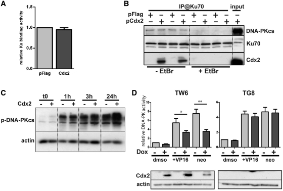
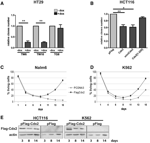
Cdx2, a gene of the paraHox cluster, encodes a homeodomain transcription factor that plays numerous roles in embryonic development and in homeostasis of the adult intestine. Whereas Cdx2 exerts a tumor suppressor function in the gut, its abnormal ectopic expression in acute leukemia is associated to a pro-oncogenic function. To try to understand this duality, we have hypothesized that Cdx2 may interact with different protein partners in the two tissues and set up experiments to identify them by tandem affinity purification. We show here that Cdx2 interacts with the Ku heterodimer specifically in intestinal cells, but not in leukemia cells, via its homeodomain. Ku proteins do not affect Cdx2 transcriptional activity. However, Cdx2 inhibits in vivo and in vitro the DNA repair activity mediated by Ku proteins in intestinal cells. Whereas Cdx2 does not affect the recruitment of Ku proteins and DNA-PKcs into the DNA repair complex, it inhibits DNA-PKcs activity. Thus, we report here a new function of Cdx2, acting as an inhibitor of the DNA repair machinery, that may contribute to its tumor suppressor function specifically in the gut.


Figures

Figure 1.
Figure 1.
Cdx2 interacts with Ku70/Ku80 proteins. (A) Silver-stained protein gel obtained after tandem affinity purification from K562, Nalm6, HCT116 and SW480 cells transfected with pCTAP-Cdx2. M: molecular weight protein ladder. Cdx2-CBP-SBP (Cdx2-cs) and Ku70/Ku80 proteins are indicated with gray or black arrowheads, respectively. (B) Western blot revealing the presence of Ku70 and Ku80 proteins in all studied cell lines. Nearly 20 µg of total proteins were separated on a gradient SDS–PAGE. Actin was used as loading control. (C) Coimmunoprecipitation of endogenous Cdx2 and Ku proteins. SW480 extracts were prepared and Cdx2, Ku70 or Ku80 were immunoprecipitated using corresponding antibodies. Mouse IGg (M-IGg) or rabbit IGg (R-IGg) were used as negative control. Western blot was revealed using rabbit anti-Ku70 and rabbit anti-Ku80 in the upper panel and using mouse anti-Cdx2 in the lower panel. About 20 µg of protein extracts were loaded in the input line. (D) In situ proximity ligation assay. Mouse anti-Cdx2 and rabbit anti-Ku70 (left panel) or mouse anti-Cdx2 and rabbit anti-Ku80 antibodies (right panel) were used to reveal endogenous proteins in SW480 cells. Green fluorescence corresponds to the PLA positive signal and indicates that the two molecules belong to the same protein complex; blue fluorescence corresponds to nuclei (DAPI staining). Red bar is 10 µm.
Figure 2.
Figure 2.
The homeodomain of Cdx2 is necessary for its interaction with Ku70/Ku80. (A) Schematic representation of the different Cdx2 deletion mutants tested. TAD: transactivator domain; NLS: nuclear localization signal; HD: homeodomain; SD: stabilization domain. The upper numbers represents the position of the different domains in the 313aa long Cdx2 protein. (B) Coimmunoprecipitations of the different mutant proteins with Ku proteins. Co-IP was performed in HCT116 transfected with the indicated mutant plasmids using anti-Flag antibodies. Immunoprecipitates were separated on a SDS–PAGE and analyzed by western blot using anti-Ku70 and anti-Ku80 antibodies (upper panel), anti-Flag antibodies (middle panel). Ku70 and Ku80 proteins present in the protein extracts before immunoprecipitation were revealed by western blot (input, lower panel).
Figure 3.
Figure 3.
Ku proteins do not affect significantly Cdx2 transcriptional activity. (A) Stimulation of endogenous Cdx2 target genes. HCT116 were transfected with 1 µg of each plasmid in a combined manner as indicated and RNA were extracted and analyzed by RT-qPCR using CDH17, SI or TBP TaqMan probes. Results are represented as fold induction relative to pFlag (normalized with TBP expression). Data correspond to the mean of several experiments and error bars represent SEM (n = 2). (B) Stimulation of Cdx2 reporter plasmids. HCT116 were transfected with CDH17-luc or SI-luc reporter plasmids and with indicated expression plasmids. Protein extracts were analyzed for dual luciferase activity. Relative promoter activity (normalized with Renilla values) of reporter plasmid cotransfected with empty plasmids was set at 1. Data correspond to the mean of several experiments and error bars represent SEM (n = 3). Right panel: FOXO4 transcription factor was used as positive control for Ku70 inhibition on the 6DBE-luc reporter plasmid as described in Brenkman et al. (27).
Figure 4.
Figure 4.
Cdx2 inhibits in vitro DNA repair in intestinal cells but not in leukemia cells. (A) NHEJ-C predominates in HCT116 cells. Linear DNA was incubated with nuclear extracts of HCT116 cells transfected with the siRNA against Ku70 or control siRNA. Processed plasmids were separated from the linear DNA by gel electrophoresis. When indicated, wortmanin or anti-PARP-1 antibodies were added to the reaction. (B) Cdx2 inhibits in vitro DNA repair in HCT116 cells. HCT116 cells were transfected with empty vector or pFlag-Cdx2 plasmids and nuclear extracts were assayed for in vitro DNA repair. When indicated (-ATP), ATP was omitted in the reaction. (C) Cdx2[1–220] mutant does not inhibits DNA repair. HCT116 cells were transfected with empty vector or Cdx2 mutant plasmids as indicated and nuclear extracts were assayed for in vitro DNA repair. When indicated (-ATP), ATP was omitted in the reaction. Above, expression of Cdx2 and its mutants was checked by western blot on 10 µg of whole-protein extracts using anti-Flag antibody. Actin was used as loading control. (D) Cdx2 does not alter in vitro DNA repair in Nalm6 cells. Nalm6 cells were transfected with empty vector or pFlag-Cdx2 plasmids and nuclear extracts were assayed for in vitro DNA repair. Gray arrows indicate linear plasmid and black arrows multimeric forms of the plasmid.
Figure 5.
Figure 5.
Cdx2 inhibits DNA repair in vivo in intestinal cells. (A) Schematic representation of the pEGFP-Pem1-Ad2 plasmid. G and FP corresponds to the 5′ and 3′ part of the GFP encoding cDNA. Ad corresponds to the adenoviral intron flanked by two HindIII sites. pCMV indicates the CMV promoter. (B) Example of green-versus-red fluorescent plots. The experiment was performed in HCT116 transfected with pFlag (left panel) or pFlag-Cdx2 (right panel). X axis: green fluorescence; Y axis: red fluorescence. (C) Cdx2 alters the in vivo DNA repair. Results represent the mean of at least four experiments performed in HCT116 (left panel) or HT29 (right panel) transfected with either pFlag, pFlag-Cdx2, pFlag-miniCdx2 or Cdx2[1-220] plasmids as indicated. The percentage of GFP and Cherry double positive cells in the Cherry positive cell population was set at 1 for pFlag and error bars represent SEM. Asterisk indicate a significant difference (**P < 0.01; *P < 0.05). Above, expression of Cdx2 and its mutants was checked by western blot on 10 µg of whole protein extracts using anti-Flag antibody. Actin was used as loading control.
Figure 6.
Figure 6.
Cdx2 does alter neither Ku proteins and DNA-PKcs recruitment nor DNA-PK autophosphorylation but inhibits DNA-PKcs activity. (A) Cdx2 does not interfere with DSB Ku70 binding. HCT116 cells were transfected with pFlag (light gray) or Cdx2 (dark gray) expressing plasmid and Ku70 binding activity was assessed. Results represent the mean of at least four independent experiments. pFlag values were set at 1. (B) Cdx2 does not alter DNA-PKcs recruitment. HCT116 were transfected with either pFlag or pFlag-Cdx2 as indicated and immunoprecipitation was performed using anti-Ku70 antibodies in the presence or absence of Ethidium Bromide (EtBr) as indicated at the bottom of the figure. DNA-PKcs, Ku70 or Cdx2 were revealed by western blot. Twenty micrograms of whole-protein extract were loaded on the right line (input). (C) Cdx2 does not modify DNA-PKcs autophosphorylation. Time course analysis of the phosphorylation of the DNA-PKcs after etoposide treatment 100 μM for 1 hour of HCT116 cells transfected with pFlag or pFlag-Cdx2. Phospho-DNA-PKcs was revealed by western blot. β-actin was used to normalized the amount of loaded proteins. (D) Cdx2 inhibits DNA-PKcs activity. DNA-PKcs activity was assessed in HT29-TW6 or -TG8 (control) cells treated or not with etoposide (VP16) 100 μM or neocarzinostatin 200 ng/μL for 1 hour. Cdx2 expression was induced with doxycyclin when indicated (dark gray bars). Experiments without doxycyclin and etoposide treatment were considered as references and results were set at 1. Results illustrate the mean of at least six experiments and error bars represent SEM. Asterisk indicates a significant difference (**P < 0.01; *P < 0.05). Above, Cdx2 expression was checked by western blot using anti-Cdx2 antibody on 10 µg of whole-protein extracts. Actin was used as loading control.
Figure 7.
Figure 7.
Cdx2 inhibits cell survival in intestinal cells but rather enhances cell recovery in leukemia cells. (A) Cdx2 inhibits cell survival in HT29 cells. HT29-TW6, -TW10 or -TG8 were treated with etoposide in the presence (dark gray) or not (light gray) of doxycyclin and cultured for 2 weeks. (B) Cdx2 inhibits cell survival in HCT116 cells. HCT116 cells were transfected with either pFlag, pFlag-Cdx2, pFlag-miniCdx2 or pFlag-Cdx2[1-220] plasmids as indicated, treated with etoposide and cultured for 2 weeks. For A and B, the numbers of clones are represented as the mean of four independent experiments. Values corresponding to -dox for HT29 or pFlag for HCT116 were set at 1 and error bars represent SEM. Asterisks indicate a significant difference (**P < 0.01; *P < 0.05). (C) and (D) Cdx2 does not alter cell survival in leukemia cells. Nalm6 or K562 cells were transfected with vehicle or pFlag-Cdx2 and cell death number was quantified using annexinV-FITC/propidium iodide at different times after etoposide treatment as indicated. Results are represented as percentage of living cells. For C and D, results of a representative experiment performed in triplicates are shown. (E) Control of Cdx2 protein expression. At 3, 8, or 14 days after transfection with indicated plasmids, HCT116 or K562 cells were lysed in laemmli buffer and Cdx2 expression was tested by western blot on 10 µg of whole protein extracts using anti-Flag antibody. Actin was used as loading control.

Similar articles

Cited by

References

    1. Chawengsaksophak K, James R, Hammond VE, Kontgen F, Beck F. Homeosis and intestinal tumours in Cdx2 mutant mice. Nature. 1997;386:84–87. - PubMed
    1. Chawengsaksophak K, De Graaff W, Rossant J, Deschamps J, Beck F. Cdx2 is essential for axial elongation in mouse development. Proc. Natl Acad. Sci. USA. 2004;101:7641–7645. - PMC - PubMed
    1. Van Den Akker E, Forlani S, Chawengsaksophak K, De Graaff W, Beck F, Meyer BI, Deschamps J. Cdx1 and Cdx2 have overlapping functions in anteroposterior patterning and posterior axis elongation. Development. 2002;129:2181–2193. - PubMed
    1. Beck F, Chawengsaksophak K, Waring P, Playford RJ, Furness JB. Reprogramming of intestinal differentiation and intercalary regeneration in Cdx2 mutant mice. Proc. Natl Acad. Sci. USA. 1999;96:7318–7323. - PMC - PubMed
    1. Gao N, White P, Kaestner KH. Establishment of intestinal identity and epithelial-mesenchymal signaling by Cdx2. Dev. Cell. 2009;16:588–599. - PMC - PubMed

Publication types

MeSH terms