

Differentiation of the seven major lyssavirus species by oligonucleotide microarray
- PMID: 22189108
- PMCID: PMC3295130
- DOI: 10.1128/JCM.00848-11
Differentiation of the seven major lyssavirus species by oligonucleotide microarray
Abstract
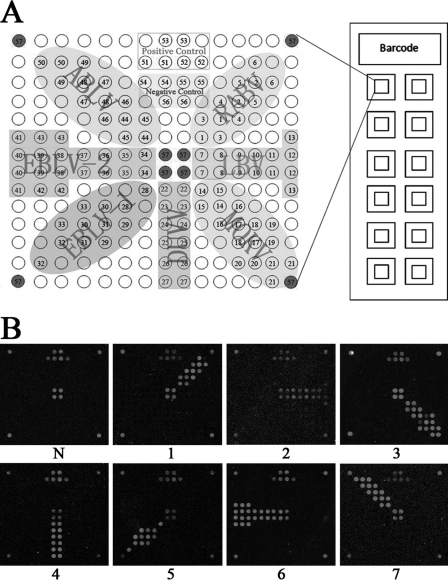
An oligonucleotide microarray, LyssaChip, has been developed and verified as a highly specific diagnostic tool for differentiation of the 7 major lyssavirus species. As with conventional typing microarray methods, the LyssaChip relies on sequence differences in the 371-nucleotide region coding for the nucleoprotein. This region was amplified using nested reverse transcription-PCR primers that bind to the 7 major lyssaviruses. The LyssaChip includes 57 pairs of species typing and corresponding control oligonucleotide probes (oligoprobes) immobilized on glass slides, and it can analyze 12 samples on a single slide within 8 h. Analysis of 111 clinical brain specimens (65 from animals with suspected rabies submitted to the laboratory and 46 of butchered dog brain tissues collected from restaurants) showed that the chip method was 100% sensitive and highly consistent with the "gold standard," a fluorescent antibody test (FAT). The chip method could detect rabies virus in highly decayed brain tissues, whereas the FAT did not, and therefore the chip test may be more applicable to highly decayed brain tissues than the FAT. LyssaChip may provide a convenient and inexpensive alternative for diagnosis and differentiation of rabies and rabies-related diseases.
Figures
Similar articles
-
A Pan-Lyssavirus Taqman Real-Time RT-PCR Assay for the Detection of Highly Variable Rabies virus and Other Lyssaviruses.PLoS Negl Trop Dis. 2017 Jan 12;11(1):e0005258. doi: 10.1371/journal.pntd.0005258. eCollection 2017 Jan. PLoS Negl Trop Dis. 2017. PMID: 28081126 Free PMC article.
-
Development of a DNA microarray for simultaneous detection and genotyping of lyssaviruses.Virus Res. 2009 Sep;144(1-2):202-8. doi: 10.1016/j.virusres.2009.04.028. Epub 2009 May 9. Virus Res. 2009. PMID: 19433118
-
Improved PCR methods for detection of African rabies and rabies-related lyssaviruses.J Clin Microbiol. 2010 Nov;48(11):3949-55. doi: 10.1128/JCM.01256-10. Epub 2010 Sep 1. J Clin Microbiol. 2010. PMID: 20810772 Free PMC article.
-
Molecular epidemiology of bat lyssaviruses in Europe.Zoonoses Public Health. 2013 Feb;60(1):35-45. doi: 10.1111/zph.12003. Epub 2012 Sep 3. Zoonoses Public Health. 2013. PMID: 22937876 Review.
-
Molecular epidemiology of lyssaviruses in Eurasia.Dev Biol (Basel). 2008;131:125-31. Dev Biol (Basel). 2008. PMID: 18634471 Review.
Cited by
-
Rabies in Costa Rica - Next Steps Towards Controlling Bat-Borne Rabies After its Elimination in Dogs.Yale J Biol Med. 2021 Jun 30;94(2):311-329. eCollection 2021 Jun. Yale J Biol Med. 2021. PMID: 34211351 Free PMC article. Review.
References
-
- Amengual B, Whitby JE, King A, Cobo JS, Bourhy H. 1997. Evolution of European bat lyssaviruses. J. Gen. Virol. 78:2319–2328 - PubMed
-
- Baxi MK, Baxi S, Clavijo A, Burton KM, Deregt D. 2006. Microarray-based detection and typing of foot-and-mouth disease virus. Vet. J. 172:473–481 - PubMed
-
- Berthet N, et al. 2010. High-density resequencing DNA microarrays in public health emergencies. Nat. Biotechnol. 28:25–27 - PubMed
Publication types
MeSH terms
Substances
LinkOut - more resources
Full Text Sources
Other Literature Sources
Research Materials
