. 2011 Dec 21:8:179.
doi: 10.1186/1742-2094-8-179.

Insulin-like growth factor-I peptides act centrally to decrease depression-like behavior of mice treated intraperitoneally with lipopolysaccharide

Affiliations

Insulin-like growth factor-I peptides act centrally to decrease depression-like behavior of mice treated intraperitoneally with lipopolysaccharide

Sook-Eun Park et al. J Neuroinflammation. .

Abstract

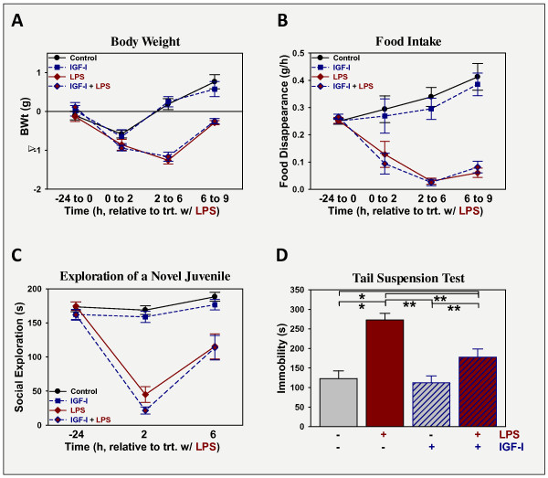
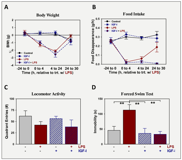
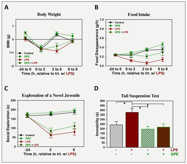
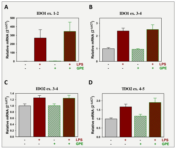
Centrally administered insulin-like growth factor (IGF)-I has anti-depressant activity in several rodent models, including lipopolysaccharide (LPS)-induced depression. In this study we tested the ability of IGF-I and GPE (the N-terminal tri-peptide derived from IGF-I) to alter depression-like behavior induced by intraperitoneal (i.p.) administration of LPS in a preventive and curative manner. In the first case, IGF-I (1 μg) or GPE (5 μg) was administered i.c.v. to CD-1 mice followed 30 min later by 330 μg/kg body weight i.p. LPS. In the second case, 830 μg/kg body weight LPS was given 24 h prior to either IGF-I or GPE. When administered i.p., LPS induced full-blown sickness assessed as a loss of body weight, decrease in food intake and sickness behavior. None of these indices were affected by IGF-I or GPE. LPS also induced depression-like behavior; assessed as an increased duration of immobility in the tail suspension and forced swim tests. When administered before or after LPS, IGF-I and GPE abrogated the LPS response; attenuating induction of depression-like behaviors and blocking preexistent depression-like behaviors. Similar to previous work with IGF-I, GPE decreased brain expression of cytokines in response to LPS although unlike IGF-I, GPE did not induce the expression of brain-derived neurotrophic factor (BDNF). LPS induced expression of tryptophan dioxygenases, IDO1, IDO2 and TDO2, but expression of these enzymes was not altered by GPE. Thus, both IGF-I and GPE elicit specific improvement in depression-like behavior independent of sickness, an action that could be due to their anti-inflammatory properties.


Figures

Figure 1
Figure 1
IGF-I does not affect sickness, but prevents the development of depression-like behavior induced by LPS. Mice were treated with vehicle or IGF-I (1 μg i.c.v.) followed 30 min later by vehicle or LPS (330 μg/kg body weight i.p.). Sickness was assessed as changes in body weight (A) and food intake (B), while sickness behavior was assessed as change in time spent investigating a novel juvenile (C). Depression-like behavior was quantified as the time of immobility during the FST, 9 h after treatment with LPS (D). n = 17-21 per treatment.
Figure 2
Figure 2
IGF-I does not affect sickness, but relieves depression-like behavior induced by LPS. Mice were treated with vehicle or LPS (830 μg/kg body weight i.p.) followed 24 h later by vehicle or IGF-I (1 μg i.c.v.). Sickness was assessed as changes in body weight (A) and food intake (B) while sickness behavior was assessed as change in general activity, 27 h after treatment with LPS (C). Depression-like behavior was quantified as the time of immobility during the FST, 30 h after treatment with LPS (D). n = 6.
Figure 3
Figure 3
GPE does not affect sickness, but prevents the development of depression-like behavior induced by LPS. Mice were treated with vehicle or GPE (5 μg i.c.v.) followed 30 min later by vehicle or LPS (330 μg/kg body weight i.p.). Sickness was assessed as changes in body weight (A) and food intake (B), while sickness behavior was assessed as change in time spent investigating a novel juvenile (C). Depression-like behavior was quantified as the time of immobility during the FST, 9 h after treatment with LPS (D). n = 7-8.
Figure 4
Figure 4
GPE does not affect sickness, but relieves depression-like behavior induced by LPS. Mice were treated with vehicle or LPS (830 μg/kg body weight i.p.) followed 24 h later by vehicle or GPE (5 μg i.c.v.). Sickness was assessed as changes in body weight (A) and food intake (B) while sickness behavior was assessed as change in general activity, 27 h after treatment with LPS (C). Depression-like behavior was quantified as the time of immobility during the FST, 30 h after treatment with LPS, or sucrose preference (D). n = 5-6.
Figure 5
Figure 5
GPE blocks the expression of inflammatory markers that are induced by LPS. Mice were treated with vehicle or GPE (5 μg i.c.v.) followed 30 min later by vehicle or LPS (330 μg/kg body weight i.p.). Mice were sacrificed, perfused and brains collected 9 h later. Steady-state mRNA expression of pro-inflammatory markers, IL-1β (A), TNFα (B), YM-1 (C) and iNOS (D), was quantified by real-time rtPCR in the prefrontal cortex and expressed relative to GAPDH. GAPDH expression was constant across treatment, p > 0.5, and changes in gene expression shown in Figures 5, 6, and 7 reflect treatment-induced effects on target gene expression. n = 11-12.
Figure 6
Figure 6
LPS increases the expression of tryptophan dioxygenases, but GPE does not attenuate this response. Mice were treated as in Figure 5. Steady-state mRNA expression, of three enzymes that metabolize tryptophan to kynurenine, IDO1 (A, B), IDO2 (C) and TDO2 (D), was quantified by real-time rtPCR in the prefrontal cortex and expressed relative to GAPDH. n = 11-12.
Figure 7
Figure 7
LPS depresses the expression of neurotrophic factors, but GPE does not alter the LPS response. Mice were treated as in Figure 5. Steady-state mRNA expression of several neurotrophic factors, IGF-I (A), VEGF (B) and BDNF (C, D), was quantified by real-time rtPCR in the prefrontal cortex and expressed relative to GAPDH. n = 11-12.

Similar articles

Cited by

References

    1. Raison CL, Capuron L, Miller AH. Cytokines sing the blues: inflammation and the pathogenesis of depression. Trends Immunol. 2006;27:24–31. doi: 10.1016/j.it.2005.11.006. - DOI - PMC - PubMed
    1. Miller AH, Maletic V, Raison CL. Inflammation and its discontents: the role of cytokines in the pathophysiology of major depression. Biol Psychiatry. 2009;65:732–741. doi: 10.1016/j.biopsych.2008.11.029. - DOI - PMC - PubMed
    1. Dantzer R, O'Connor JC, Freund GG, Johnson RW, Kelley KW. From inflammation to sickness and depression: when the immune system subjugates the brain. Nat Rev Neurosci. 2008;9:46–56. doi: 10.1038/nrn2297. - DOI - PMC - PubMed
    1. Reichenberg A, Yirmiya R, Schuld A, Kraus T, Haack M, Morag A, Pollmacher T. Cytokine-associated emotional and cognitive disturbances in humans. Arch Gen Psychiatry. 2001;58:445–452. doi: 10.1001/archpsyc.58.5.445. - DOI - PubMed
    1. Eisenberger NI, Berkman ET, Inagaki TK, Rameson LT, Mashal NM, Irwin MR. Inflammation-induced anhedonia: endotoxin reduces ventral striatum responses to reward. Biol Psychiatry. 2010;68:748–754. doi: 10.1016/j.biopsych.2010.06.010. - DOI - PMC - PubMed

Publication types

MeSH terms