. 2012 Apr;37(5):1305-20.
doi: 10.1038/npp.2011.319. Epub 2011 Dec 21.

Gap junction dysfunction in the prefrontal cortex induces depressive-like behaviors in rats

Affiliations

Gap junction dysfunction in the prefrontal cortex induces depressive-like behaviors in rats

Jian-Dong Sun et al. Neuropsychopharmacology. 2012 Apr.

Abstract

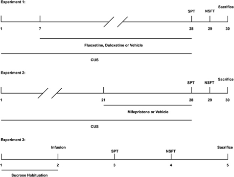
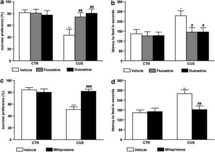
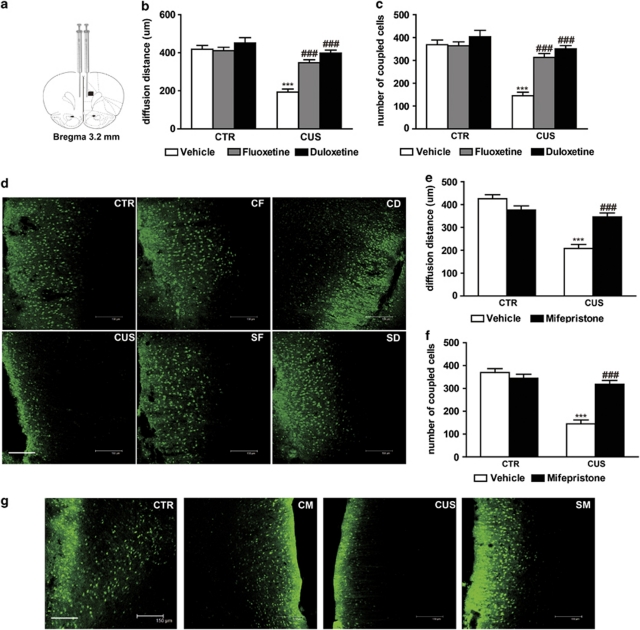
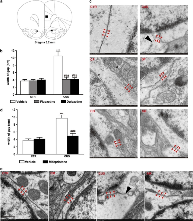
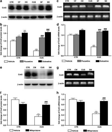
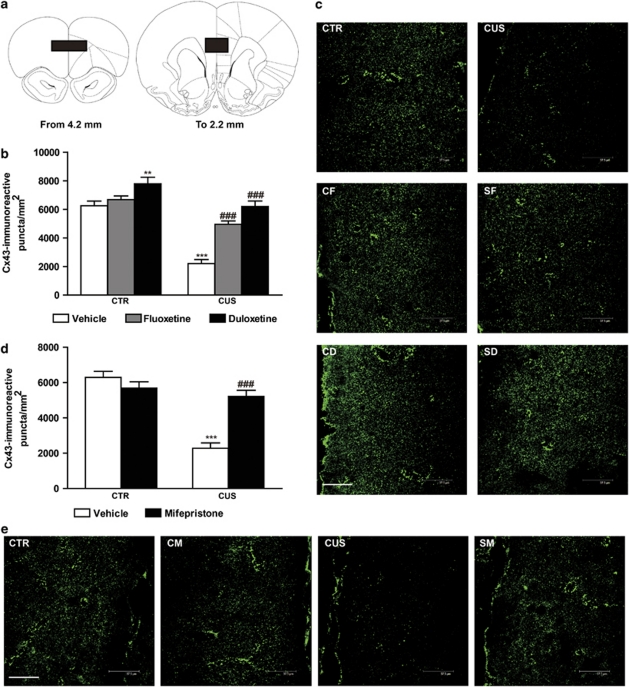
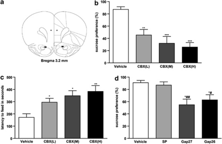
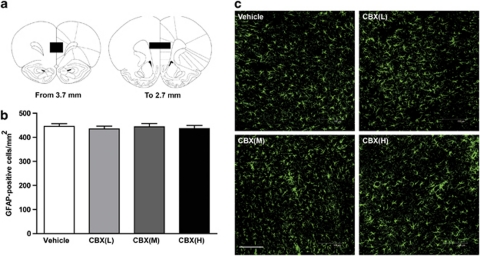
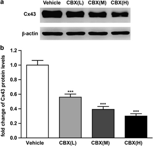
Growing evidence has implicated glial anomalies in the pathophysiology of major depression disorder (MDD). Gap junctional communication is a main determinant of astrocytic function. However, it is unclear whether gap junction dysfunction is involved in MDD development. This study investigates changes in the function of astrocyte gap junction occurring in the rat prefrontal cortex (PFC) after chronic unpredictable stress (CUS), a rodent model of depression. Animals exposed to CUS and showing behavioral deficits in sucrose preference test (SPT) and novelty suppressed feeding test (NSFT) exhibited significant decreases in diffusion of gap junction channel-permeable dye and expression of connexin 43 (Cx43), a major component of astrocyte gap junction, and abnormal gap junctional ultrastructure in the PFC. Furthermore, we analyzed the effects of typical antidepressants fluoxetine and duloxetine and glucocorticoid receptor (GR) antagonist mifepristone on CUS-induced gap junctional dysfunction and depressive-like behaviors. The cellular and behavioral alterations induced by CUS were reversed and/or blocked by treatment with typical antidepressants or mifepristone, indicating that the mechanism of their antidepressant action may involve the amelioration of gap junction dysfunction and the cellular changes may be related to GR activation. We then investigated the effects of pharmacological gap junction blockade in the PFC on depressive-like behaviors. The results demonstrate that carbenoxolone (CBX) infusions induced anhedonia in SPT, and anxiety in NSFT, and Cx43 mimetic peptides Gap27 and Gap26 also induced anhedonia, a core symptom of depression. Together, this study supports the hypothesis that gap junction dysfunction contributes to the pathophysiology of depression.


Figures

Figure 1
Figure 1
Experimental design. In experiment 1, rats were handled daily (home cage control, CTR) or subjected to the chronic unpredictable stress (CUS) procedure for 28 days. Animals were administered vehicle, fluoxetine (10 mg/kg), or duloxetine (10 mg/kg) for the 21 last days of the experiment. The efficacy of CUS or antidepressants on behavioral performances of the animals in sucrose preference test (SPT) and novelty suppressed feeding test (NSFT) was measured. In experiment 2, rats were handled daily (home cage control, CTR) or subjected to the CUS procedure for 28 days. Animals were administered vehicle or mifepristone (50 mg/kg) for the 7 last days of the experiment. SPT and NSFT were also performed. In experiment 3, animals were infused with 1, 10, and 100 mM carbenoxolone (CBX) or with scrambled peptides, Gap27 and Gap26, into the prefrontal cortex (PFC), and were then subjected to the SPT and NSFT. Phosphate-buffered saline (PBS, pH 7.4) was the vehicle in the control group.
Figure 2
Figure 2
Antidepressants and mifepristone reversed behavioral deficits induced by chronic unpredictable stress (CUS; n=12/group). (a) Sucrose preference was decreased by 28 days of CUS exposure and was reversed by antidepressants treatment. (b) CUS animals showed a significant increase of latency to feed and this effect was reversed by antidepressants treatment. (c) Sucrose preference was decreased by 28 days of CUS exposure and was reversed by mifepristone treatment. (d) CUS animals showed a significant increase of latency to feed and this effect was reversed by mifepristone treatment. Error bars represent SEM. *P<0.05, **P<0.01, ***P<0.001 compared with CTR and #P<0.05, ##P<0.01, ###P<0.001 compared with CUS, two-way analysis of the variance (ANOVA), Newman–Keuls post-hoc analysis.
Figure 3
Figure 3
Antidepressants and mifepristone reversed intercellular diffusion decrease of Lucifer yellow in the prelimbic cortex (PLC) induced by chronic unpredictable stress (CUS; n=6/group). (a) The infusion illustration is shown and the distance of diffusion and the number of coupled cells in the black square are quantified (Paxinos and Watson, 1998). (b, e) The diffusion distance was significantly reduced by CUS and this effect was reversed by antidepressants or mifepristone treatment. (c, f) CUS significantly decreased the number of coupled cells and antidepressants or mifepristone significantly reversed this effect. Error bars represent SEM. ***P<0.001 compared with CTR and ###P<0.001 compared with CUS, two-way analysis of the variance (ANOVA), Newman–Keuls post-hoc analysis. (d, g) Representative dye coupling images from animals of all groups are illustrated. Scale bar=150 μm. CTR, control group; CUS, CUS group; CF, CTR+fluoxetine group; SF, CUS+fluoxetine group; CD, CTR+duloxetine group; SD, CUS+duloxetine group; CM, CTR+mifepristone group; SM, CUS+mifepristone group.
Figure 4
Figure 4
Antidepressants and mifepristone reversed ultrastructural alterations of astrocyte gap junction in the prelimbic cortex (PLC) induced by chronic unpredictable stress (CUS; n=6/group). (a) The sections shown in the black square were cut from the brains of all groups (Paxinos and Watson, 1998). (b, d) The width of gap was significantly enlarged by CUS and this effect was reversed by antidepressants and mifepristone treatment. Error bars represent SEM. ***P<0.001 compared with CTR and ###P<0.001 compared with CUS, two-way analysis of the variance (ANOVA), Newman–Keuls post-hoc analysis. (c, e) Electron micrographs showing astrocytic gap junction in the PLC of each group (Magnification: × 100 000). Astrocytic gap junctions are indicated by gray arrowheads. Black arrowhead indicates the damaged structure between two adjacent astrocytes. Scale bar=500 nm. CTR, control group; CUS, CUS group; CF, CTR+fluoxetine group; SF, CUS+fluoxetine group; CD, CTR+duloxetine group; SD, CUS+duloxetine group; CM, CTR+mifepristone group; SM, CUS+mifepristone group.
Figure 5
Figure 5
Antidepressants and mifepristone reversed decreases of connexin 43 (Cx43) protein and mRNA levels in the prefrontal cortex (PFC) induced by chronic unpredictable stress (CUS; n=6/group). The protein levels of Cx43 were determined by western blot. (a, e) Representative western blot images of Cx43 and β-actin of each group are shown, and (b, f) results were quantified and are the mean±SEM, taking the ratio of the gray level of Cx43 band to the β-actin band of control group as unity. CUS significantly decreased the protein levels of Cx43 and antidepressants or mifepristone significantly reversed this effect. The mRNA levels of Cx43 were determined by RT-PCR. (c, g) Representative gel patterns of Cx43 and β-actin PCR product bands of each group are shown. (d, h) Relative amount (mean±SEM) of Cx43 mRNA is shown, as percent of CTR. CUS significantly decreased the mRNA levels of Cx43 and antidepressants and mifepristone significantly reversed this effect. *P<0.05, **P<0.01, ***P<0.001 compared with CTR and ###P<0.001 compared with CUS, two-way analysis of the variance (ANOVA), Newman–Keuls post-hoc analysis. RT-PCR, reverse transcriptase-PCR; CTR, control group; CUS, CUS group; CF, CTR+fluoxetine group; SF, CUS+fluoxetine group; CD, CTR+duloxetine group; SD, CUS+duloxetine group; CM, CTR+mifepristone group; SM, CUS+mifepristone group.
Figure 6
Figure 6
Antidepressants and mifepristone reversed decrease of the number of connexin 43 (Cx43)-immunoreactive puncta in the prelimbic cortex (PLC) induced by chronic unpredictable stress (CUS; n=6/group). Numbers of Cx43-immunoreactive puncta were quantified throughout the PLC as shown in (a) (black squares) (Paxinos and Watson, 1998). (b, d) The number of Cx43-immunoreactive puncta/mm2 was significantly decreased by CUS and this effect was reversed by antidepressants or mifepristone treatment. Error bars represent SEM. **P<0.01, ***P<0.001 compared with CTR and ###P<0.001 compared with CUS, two-way analysis of the variance (ANOVA), Newman–Keuls post-hoc analysis. (c, e) Representative Cx43-stained sections from animals of all groups are illustrated at ∼3.2 mm from bregma. Scale bar=37.5 μm. CTR, control group; CUS, CUS group; CF, CTR+fluoxetine group; SF, CUS+fluoxetine group; CD, CTR+duloxetine group; SD, CUS+duloxetine group; CM, CTR+mifepristone group; SM, CUS+mifepristone group.
Figure 7
Figure 7
Gap junction blockade in the prefrontal cortex (PFC) induces depressive-like behaviors. (a) The infusion site (coordinates (mm): anteroposterior +3.2, dorsolateral –0.5, depth –4 from Bregma) is marked by an asterisk (*) (Paxinos and Watson, 1998). (b, c) Effects of carbenoxolone (CBX) infusions on sucrose preference test (SPT) and novelty suppressed feeding test (NSFT; n=6/group). Animals received infusions of phosphate-buffered saline (PBS, Vehicle), 1 mM of CBX (low-dose CBX, CBX(L)), 10 mM of CBX (medium-dose CBX, CBX(M)), or 100 mM of CBX (high-dose CBX, CBX(H)) and were consecutively tested in SPT (day 3) and NSFT (day 4). Results are expressed as mean±SEM ratio of sucrose solution vs total liquid consumed (b) and latency to feed in seconds (c). *P<0.05, **P<0.01, ***P<0.001 compared with Vehicle. (d) Effects of connexin 43 (Cx43) mimetic peptide Gap27 and Gap26 infusions on SPT (n=6/group). PBS (Vehicle), scrambled peptide (SP), Gap27, or Gap26 were bilaterally infused and sucrose preference was measured. *P<0.05 compared with SP and #P<0.05, ##P<0.01 compared with Vehicle.
Figure 8
Figure 8
Influence of carbenoxolone (CBX) infusions in the prefrontal cortex (PFC) on density of glial fibrillary acidic protein (GFAP)-positive cells (n=6/group). (a) Anterior-to-posterior stereotaxic coordinates of representative coronal brain slices are shown (Paxinos and Watson, 1998). Numbers of GFAP-positive cells were quantified in the prelimbic cortex (PLC) as shown in (a) (black squares). (b) Results are presented as mean±SEM, and there was no significant difference in all groups in the number of GFAP-positive cells/mm2 (one-way analysis of the variance (ANOVA), F3, 20=0.1988, P=0.896). (c) Representative GFAP immunofluorescence-stained sections from each group are shown at ∼3.7 mm from bregma, away from the infusion site. Scale bar=150 μm. Vehicle, PBS-infused group; CBX(L), low-dose (1 mM) CBX-infused group; CBX(M), medium-dose (10 mM) CBX-infused group; CBX(H), high-dose (100 mM) CBX-infused group.
Figure 9
Figure 9
Effects of carbenoxolone (CBX) infusions in the prefrontal cortex (PFC) on the protein level of connexin 43 (Cx43; n=6/group). The protein levels of Cx43 in the PFC of each group were determined. (a) Representative western blot images of Cx43 and β-actin are shown, and (b) results were quantified and are the mean±SEM, taking the ratio of the gray level of Cx43 band to the β-actin band of Vehicle as unity. ***P<0.001 compared with Vehicle. Vehicle, PBS-infused group; CBX(L), low-dose (1 mM) CBX-infused group; CBX(M), medium-dose (10 mM) CBX-infused group; CBX(H), high-dose (100 mM) CBX-infused group.

References

    1. Araque A, Parpura V, Sanzgiri RP, Haydon PG. Tripartite synapses: glia, the unacknowledged partner. Trends Neurosci. 1999;22:208–215. - PubMed
    1. Ayensu WK, Pucilowski O, Mason GA, Overstreet DH, Rezvani AH, Janowsky DS. Effects of chronic mild stress on serum complement activity, saccharin preference, and corticosterone levels in Flinders lines of rats. Physiol Behav. 1995;57:165–169. - PubMed
    1. Banasr M, Chowdhury GM, Terwilliger R, Newton SS, Duman RS, Behar KL, et al. Glial pathology in an animal model of depression: reversal of stress-induced cellular, metabolic and behavioral deficits by the glutamate-modulating drug riluzole. Mol Psychiatry. 2010;15:501–511. - PMC - PubMed
    1. Banasr M, Duman RS. Glial loss in the prefrontal cortex is sufficient to induce depressive-like behaviors. Biol Psychiatry. 2008;64:863–870. - PMC - PubMed
    1. Banasr M, Valentine GW, Li XY, Gourley SL, Taylor JR, Duman RS. Chronic unpredictable stress decreases cell proliferation in the cerebral cortex of the adult rat. Biol Psychiatry. 2007;62:496–504. - PubMed

Publication types

MeSH terms