. 2012 Feb 1;30(6):1038-49.
doi: 10.1016/j.vaccine.2011.12.044. Epub 2011 Dec 18.

In vitro culture medium influences the vaccine efficacy of Mycobacterium bovis BCG

Affiliations

In vitro culture medium influences the vaccine efficacy of Mycobacterium bovis BCG

Manjunatha M Venkataswamy et al. Vaccine. .

Abstract

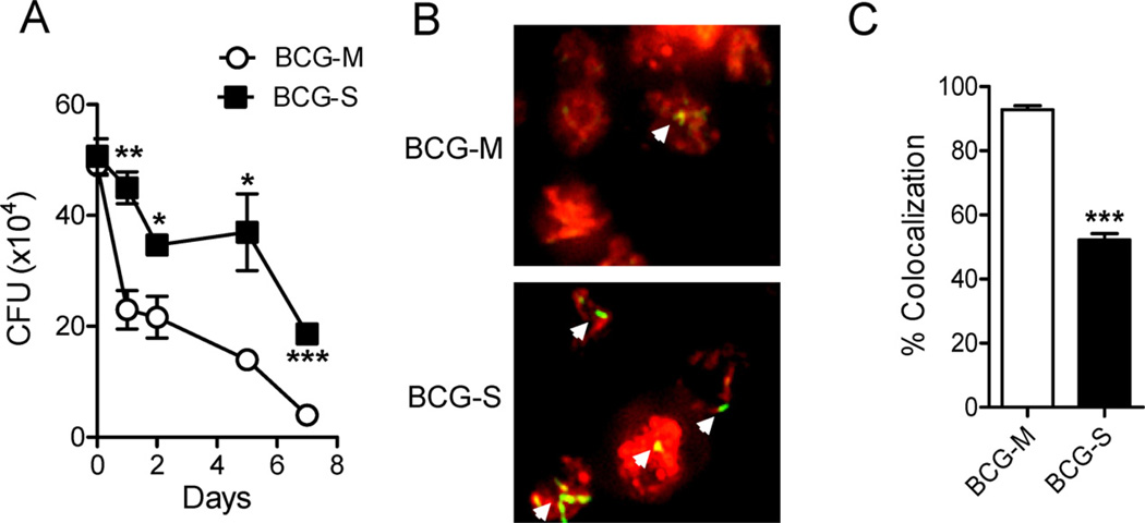
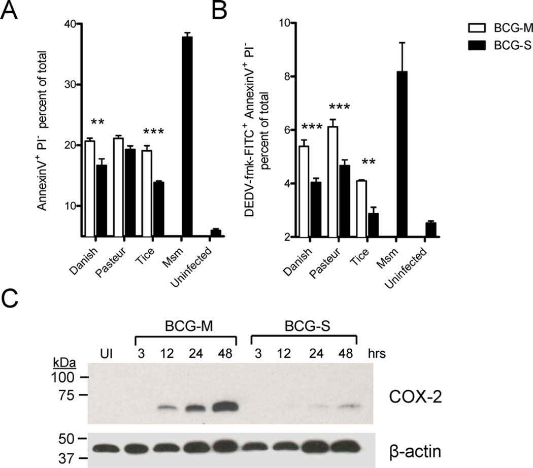
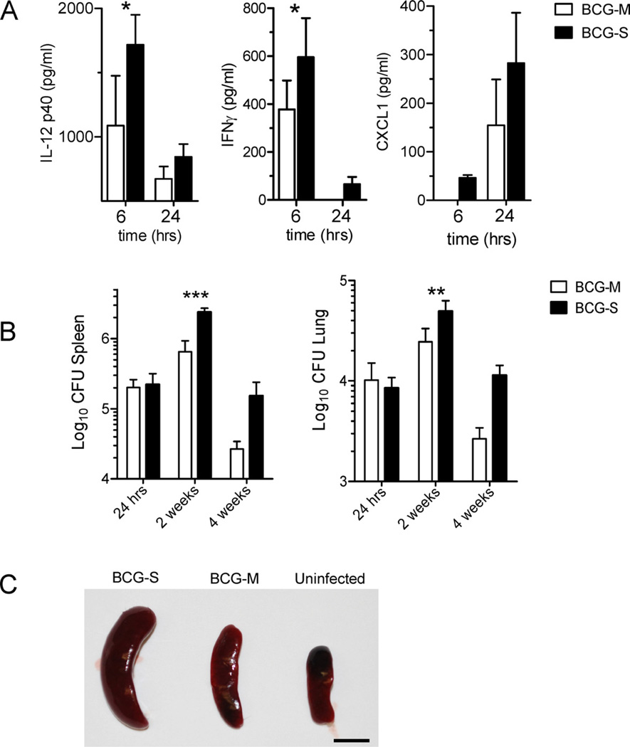
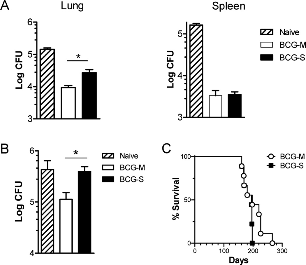
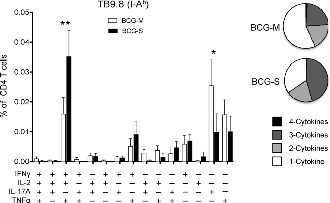
The varied rates of protection induced by Mycobacterium bovis BCG vaccine against tuberculosis has been attributed to many factors such as genetic variability among BCG strains, rapid clearance of BCG in some populations, and different levels of previous exposure of vaccinated populations to environmental mycobacteria. However, the methods and conditions employed to prepare this vaccine for human usage by various manufacturers have not been investigated as potential factors contributing to the variation in vaccine efficacy. A review of the literature indicates discrepancies between the approach for growing BCG vaccine in the laboratory to assess immune responses and protective ability in animal models, and that employed for production of the vaccine for administration to humans. One of the major differences is in the growth medium used for routine propagation in the laboratory and the one used for bulk vaccine production by manufacturers. Here we compared the immunogenicity of the BCG vaccine grown in Middlebrook 7H9 medium, the most commonly used medium in laboratory studies, against that grown in Sauton medium, which is used for growing BCG by most manufacturers. Our results showed clear differences in the behavior of BCG grown in these different culture media. Compared to BCG grown in Middlebrook 7H9 medium, BCG grown in Sauton media was more persistent inside macrophages, more effective at inhibiting apoptosis of infected cells, induced stronger inflammatory responses and stimulated less effective immunity against aerosol challenge with a virulent Mtb strain. These findings suggested that the growth medium used for producing BCG vaccine is an important factor that deserves increased scrutiny in ongoing efforts to produce more consistently effective vaccines against Mtb.


Figures

Fig. 1
Fig. 1
In vitro survival of BCG-M and BCG-S in BMM. (A) Bone marrow derived macrophages were infected at an MOI of 10:1, and CFU counts were obtained at various time points after infection. Data shown are representative of 3 independent experiments (*p < 0.05, **p < 0.01, ***p < 0.005). (B) Bone marrow derived macrophages were infected with BCG-M or BCG-S labeled with CFSE (indicated by green or yellow color) and stained with Lysotracker dye (red color) at 24 h after infection, followed by analysis using fluorescence microscopy (magnification 20×). Representative images are shown from 2 independent experiments that yielded similar results. White arrows indicate CFSE-labeled bacteria. (C) Quantification of co-localization of Lysotracker with CFSE stained BCG (***p < 0.0001). Note that the Pasteur strain of BCG was used in the experiments shown in this figure, and in all subsequent figures except where otherwise state.
Fig. 2
Fig. 2
Increased apoptosis of host cells with infection by BCG-M. (A and B) THP-1 cells were infected with BCG-M or BCG-S and analyzed 72 h later by FACS for the induction of apoptosis by staining with Annexin V-Alexa Fluor® 647 and Propidium Iodide (A), and FITC-DEVD-fmk (B) (**p < 0.01, ***p < 0.001). (C) BMMs were infected and lysates obtained at various time points to measure COX-2 expression levels by Western Blot. Each lane was loaded with an equal amount of lysate comprising 3 × 10⁶ cell equivalents and normalized according to the signal for the beta-actin control. UI indicates uninfected BMMs. Results shown are representative of two experiments.
Fig. 3
Fig. 3
Increased inflammatory cytokine response and enhanced bacterial survival in vivo with BCG-S. (A) Cytokine levels in sera of mice infected i.v. with either 1 × 10⁶ BCG-M or BCG-S were analyzed by multiplex capture ELISA at 6 h and 24 h post infection. Results are shown for IL-12p40, IFNγ, and CXCL1, which were the only three cytokines in the 10-plex analysis system that showed significantly different levels betweens the two groups of mice. Bars represent means of cytokine levels from 3 mice in each group, and error bars show standard errors. (B) In vivo CFU loads of BCG in the spleens (left) and lungs (right) after intravenous infection of C57BL/6 mice with 1 × 10⁶ CFU of BCG-M or BCG-S. Results are averages from 3 mice in each group. For (A) and (B): *p < 0.05, **p < 0.01, ***p < 0.001. (C) Representative spleens are shown from C57BL/6 mice that were infected intraperitoneally with either 5 × 10⁶ CFU of BCG-S or BCG-M for 2 weeks, or from a mouse that received only saline injection (uninfected). Scale bar corresponds to 1 cm.
Fig. 4
Fig. 4
Induction of more robust T cell responses to mycobacterial antigens by BCG-S. (A) Multiparameter FACS with intracellular staining for cytokines in splenocytes from animals immunized 2 weeks previously with 5 × 10⁶ CFU of BCG-M (open bars) or BCG-S (filled bars) intraperitoneally, and restimulated in vitro with peptides corresponding to epitopes of the indicated mycobacterial antigens plus soluble anti-CD28 mAb. The graphs show the percentages of total CD8+ T cells (for TB10.3/10.4) or CD4+ T cells (for Ag85 P25 and TB9.8) producing IFNγ, IL-2 or TNFα and combinations of two or three of these cytokines; *p < 0.05, ***p < 0.001. (B) Pie charts summarizing frequencies of cells producing one, two or three cytokines in the experiment shown in A. (C) ELISPOT assay for IFNγ producing cells in splenocytes from mice immunized i.p. with 5 × 10⁶ CFU of either BCG-M or BCG-S 2 weeks earlier. The cells were stimulated in vitro with peptides corresponding to epitopes for the indicated mycobacterial antigens recognized by CD4+ T cells (Ag85B, TB9.8) or CD8+ T cells (TB10.3/4) as indicated, and with recombinant Hsp65 protein that contains epitopes for both CD4+ and CD8+ T cell responses; *p < 0.05. All results shown are means and SD for groups of 5 mice immunized with each BCG preparation.
Fig. 5
Fig. 5
Improved vaccine efficacy against M. tuberculosis challenge with BCG-M. C57BL/6 mice were vaccinated subcutaneously with 1 × 10⁶ CFU of BCG-M or BCG-S, or sham-vaccinated with saline. Two months later, animals were challenged by aerosol infection with 50–100 CFU of virulent M. tuberculosis (strain H37Rv). (A) Bars show means and SD for CFU of M. tuberculosis in lungs (left) and spleens (right) at 3 months after challenge for groups of 5 mice. Results shown are representative of two independent experiments. (B) CFU counts from lungs of mice challenged with M. tuberculosis after adoptive transfer of T cells from donor mice immunized with either BCG-M or BCG-S (1 × 10⁶ CFU subcutaneously) or sham immunized with saline (naïve) mice as indicated. Results shown are representative of two independent experiments, each with 5 mice per group. In (A) and (B), *p < 0.05 (unpaired t test). (C) Survival curves for SCID mice (N = 9 mice per group) after i.v. infection with 1 × 10⁶ BCG-M or BCG-S. Differences between the survival curves were not significant (p > 0.05, Log rank test).
Fig. 6
Fig. 6
Induction of stronger Th17 responses by BCG-M. Multiparameter FACS with intracellular staining for cytokines in splenocytes from animals immunized 8 weeks previously with 1 × 10⁶ CFU of BCG-M or BCG-S subcutaneously, and restimulated in vitro with I-Ab restricted TB9.8 peptide plus soluble anti-CD28 mAb. The bar graph shows the percentages of total CD4+ T cells producing IFNγ, IL-2, IL-17A or TNFα and combinations of two or three or four of these cytokines. Open bars correspond to BCG-M immunized mice, and filled bars are BCG-S vaccinated mice (N = 5 mice per group; *p < 0.05, **p < 0.005). Pie charts show proportions of CD4+ cells producing one, two, three or four of the cytokines analyzed following stimulation with TB9.8 peptide.
Fig. 7
Fig. 7
Distinct humoral responses induced by BCG-S and BCG-M. Pooled sera from five mice infected 2 weeks earlier with 5 × 10⁶ CFU i.p. of either BCG-M (indicated by M above relevant lanes) or BCG-S (indicated by S) were analyzed for antibody reactivity by Western blotting of M. tuberculosis proteins. Blots show reactivity with total Mtb lysate (Total), culture filtrate proteins (CFP), cell wall fraction (CW) or cytosolic fraction (Cytosol) of M. tuberculosis.

References

    1. Oettinger T, Jorgensen M, Ladefoged A, Haslov K, Andersen P. Development of the Mycobacterium bovis BCG vaccine: review of the historical and biochemical evidence for a genealogical tree. Tuberc Lung Dis. 1999;79(4):243–250. - PubMed
    1. Ritz N, Curtis N. Mapping the global use of different BCG vaccine strains. Tuberculosis (Edinb) 2009 July;89(4):248–251. - PubMed
    1. Fine PE. Variation in protection by BCG: implications of and for heterologous immunity. Lancet. 1995 November;346(8986):1339–1345. - PubMed
    1. Colditz GA, Brewer TF, Berkey CS, Wilson ME, Burdick E, Fineberg HV, et al. Efficacy of BCG vaccine in the prevention of tuberculosis. Meta-analysis of the published literature. J Am Med Assoc. 1994 March;271(9):698–702. - PubMed
    1. Russell DG, Barry CE, III, Flynn JL. Tuberculosis: what we don’t know, can, and does, hurt us. Science. 2010 May;328(5980):852–856. - PMC - PubMed

Publication types