Review
. 2011 Dec 22;2(12):e244.
doi: 10.1038/cddis.2011.130.

Cardiomyocyte death: mechanisms and translational implications

Affiliations
Review

Cardiomyocyte death: mechanisms and translational implications

M Chiong et al. Cell Death Dis. .

Abstract

Cardiovascular disease (CVD) is the leading cause of morbidity and mortality worldwide. Although treatments have improved, development of novel therapies for patients with CVD remains a major research goal. Apoptosis, necrosis, and autophagy occur in cardiac myocytes, and both gradual and acute cell death are hallmarks of cardiac pathology, including heart failure, myocardial infarction, and ischemia/reperfusion. Pharmacological and genetic inhibition of autophagy, apoptosis, or necrosis diminishes infarct size and improves cardiac function in these disorders. Here, we review recent progress in the fields of autophagy, apoptosis, and necrosis. In addition, we highlight the involvement of these mechanisms in cardiac pathology and discuss potential translational implications.


Figures

Figure 1
Figure 1
Aging, stress, and cell death progression. The cell injury marker troponin T increased during aging, as well as with different CVDs (de Lemos et al.)
Figure 2
Figure 2
Schematic overview of the regulators of autophagy described in this section: Atg5 and Beclin 1 – components of the core autophagic machinery; mTOR and Raptor are components of the mTOR complex 1, an upstream repressor of autophagy; rapamycin – an inhibitor of mTOR (activates autophagy by releasing mTOR-mediated inhibition)
Figure 3
Figure 3
The Goldilocks rule of autophagy in heart disease. Autophagy is a dynamic process. The relationship between autophagy and heart disease is complex. Although basal autophagy is critical to maintain cellular and whole-body homeostasis, both increases and decreases in autophagy to excessive degree can be maladaptive. In CH, HF, and I/R, autophagic flux is abnormally elevated, contributing to cardiac dysfunction. With aging, Pompe disease, and Danon disease, autophagic activity and processing are attenuated, perturbing cellular homeostasis and contributing to cardiac disease. Animal models have been studied extensively to evaluate the role of autophagy in heart disease with either increases (Beclin 1 tg) or decreases (Atg5 KO, Beclin 1 het) in autophagic activity
Figure 4
Figure 4
Apoptotic pathway in cardiac myocytes. In the extrinsic pathway, death receptor activation by a death ligand induces death-inducing signaling complex (DISC) formation and casp 8 activation, which in turn activates casp 3. This pathway can also activate the intrinsic pathway by the proteolysis of BID to t-BID by casp 8 and interaction of t-BID with BAX in the mitochondria. Pro-apoptotic BAX/BAK induces cyto c, Smac/DIABLO, AIF, and Endo G release from the mitochondria. Cyto c with Apaf1 and casp 9 form the apoptosome with activation of casp 9. Casp activity is regulated by the endogenous casp inhibitor XIAP. Cardiac myocytes are naturally resistant to apoptosis due to their low-level expression of Apaf1 and casps and high levels of XIAP
Figure 5
Figure 5
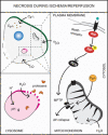
Necrosis pathways in cardiac myocytes. During certain cardiac pathologies, such as I/R, the action of cellular pumps is inhibited by ATP depletion, there is a consequent increase in H+ and Na+, and the sodium-calcium exchanger (NCX) operates in reverse manner. Increased cytoplasmic Ca2+ leads to increased Ca2+ in the mitochondrial matrix along with elevated levels of ROS, culminating in MPTP opening, and necrosis. On the other hand, mitochondrial swelling and mitochondrial membrane rupture also produce necrosis. Moreover, increased H+ in the cytoplasm and inactivation of H+ pumps elicit declines in lysosomal pH, which results in overactivation of proteases such as cathepsins. The massive entry of water results in lysosomal swelling, membrane rupture, and release of proteases into the cytoplasm, which together with other activated proteases, such as calpains, digest different substrates, including cytoskeletal proteins, contributing to necrosis. Activation of death receptors, such as the TNF-α receptor, represents other necrosis pathways in cardiac myocytes under certain conditions such as HF. The activation of these receptors could lead to the activation of receptor-interacting protein (RIP), increased ROS, and necrosis. The massive inflow of water into the cell by the osmotic imbalance ultimately leads to cell swelling and rupture of the plasma membrane

Similar articles

Cited by

References

    1. Whelan RS, Kaplinskiy V, Kitsis RN. Cell death in the pathogenesis of heart disease: mechanisms and significance. Annu Rev Physiol. 2010;72:19–44. - PubMed
    1. Levine B, Kroemer G. Autophagy in the pathogenesis of disease. Cell. 2008;132:27–42. - PMC - PubMed
    1. Marambio P, Toro B, Sanhueza C, Troncoso R, Parra V, Verdejo H, et al. Glucose deprivation causes oxidative stress and stimulates aggresome formation and autophagy in cultured cardiac myocytes. Biochim Biophys Acta. 2010;1802:509–518. - PubMed
    1. Lipinski MM, Hoffman G, Ng A, Zhou W, Py BF, Hsu E, et al. A genome-wide siRNA screen reveals multiple mTORC1 independent signaling pathways regulating autophagy under normal nutritional conditions. Dev Cell. 2010;18:1041–1052. - PMC - PubMed
    1. Matsui Y, Takagi H, Qu X, Abdellatif M, Sakoda H, Asano T, et al. Distinct roles of autophagy in the heart during ischemia and reperfusion: roles of AMP-activated protein kinase and Beclin 1 in mediating autophagy. Circ Res. 2007;100:914–922. - PubMed

Publication types