. 2011 Dec 22;2(12):e245.
doi: 10.1038/cddis.2011.131.

The role of c-FLIP splice variants in urothelial tumours

Affiliations

The role of c-FLIP splice variants in urothelial tumours

F Ewald et al. Cell Death Dis. .

Abstract

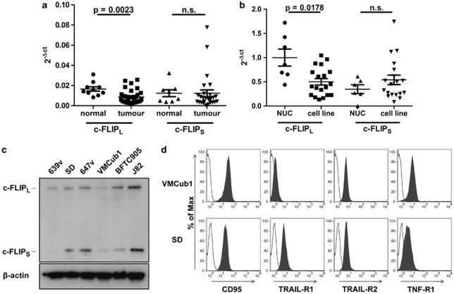
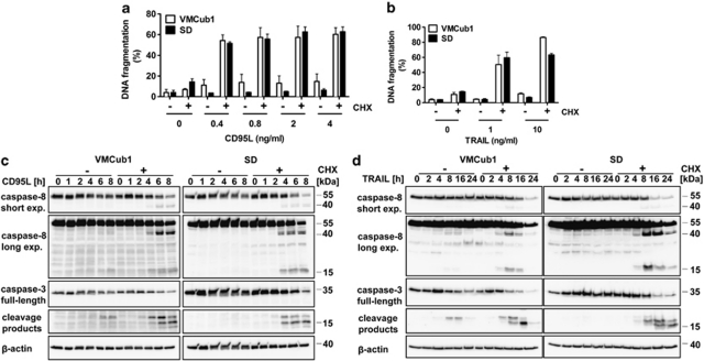
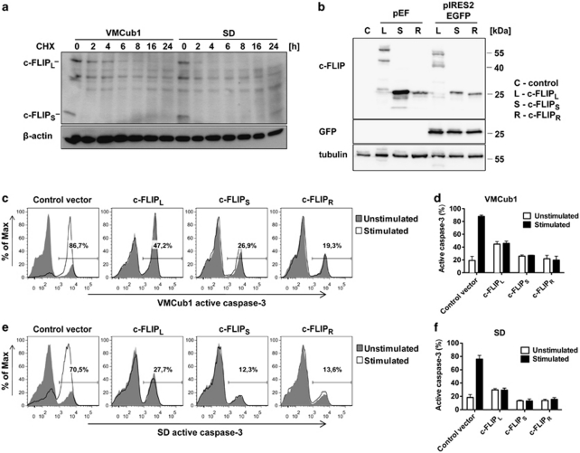
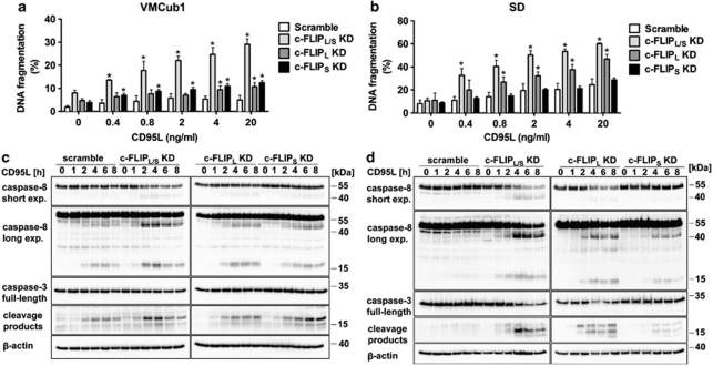
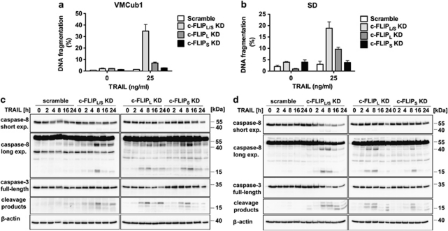
Deregulation of apoptosis is common in cancer and is often caused by overexpression of anti-apoptotic proteins in tumour cells. One important regulator of apoptosis is the cellular FLICE-inhibitory protein (c-FLIP), which is overexpressed, for example, in melanoma and Hodgkin's lymphoma cells. Here, we addressed the question whether deregulated c-FLIP expression in urothelial carcinoma impinges on the ability of death ligands to induce apoptosis. In particular, we investigated the role of the c-FLIP splice variants c-FLIP(long) (c-FLIP(L)) and c-FLIP(short) (c-FLIP(S)), which can have opposing functions. We observed diminished expression of the c-FLIP(L) isoform in urothelial carcinoma tissues as well as in established carcinoma cell lines compared with normal urothelial tissues and cells, whereas c-FLIP(S) was unchanged. Overexpression and RNA interference studies in urothelial cell lines nevertheless demonstrated that c-FLIP remained a crucial factor conferring resistance towards induction of apoptosis by death ligands CD95L and TRAIL. Isoform-specific RNA interference showed c-FLIP(L) to be of particular importance. Thus, urothelial carcinoma cells appear to fine-tune c-FLIP expression to a level sufficient for protection against activation of apoptosis by the extrinsic pathway. Therefore, targeting c-FLIP, and especially the c-FLIP(L) isoform, may facilitate apoptosis-based therapies of bladder cancer in otherwise resistant tumours.


Figures

Figure 1
Figure 1
(a) Quantification of c-FLIPL and c-FLIPS mRNA levels in normal urothelial tissues (normal; n=10) and urothelial carcinoma samples (tumour; n=28) by real-time RT-PCR. Values were normalised to GAPDH expression. Horizontal lines represent the mean; error bars display the standard deviation. Statistical analyses were performed by two-tailed Mann–Whitney U-tests. (b) Quantification of c-FLIPL and c-FLIPS mRNA levels in NUCs (n=7) and urothelial cell lines (cell line; n=19) by real-time PCR. Values were normalised to TBP expression levels. Horizontal lines represent the mean; error bars display the standard deviation. Statistical analyses were performed by two-tailed Mann–Whitney U-tests. (c) Western blot analysis of c-FLIP protein expression in the indicated urothelial cell lines. β-Actin was used as the loading control. (d) VMCub1 and SD cell surface expression of the death receptors CD95, TRAIL-R1, TRAIL-R2 and TNF-R1 analysed by flow cytometry. Unstained cells are shown in white
Figure 2
Figure 2
Urothelial carcinoma cell lines VMCub1 and SD are sensitised to apoptosis by CHX. (a and b) For analysis of apoptosis sensitivity, urothelial carcinoma cells were left untreated or stimulated for 16 h with the indicated concentrations of CD95L (a) or TRAIL (b) in the presence or absence of 10 μg/ml CHX. The amount of apoptotic cells was determined by quantification of sub-G1 DNA content by flow cytometry. Measurements are displayed as the mean of at least three independent experiments ±S.D. (c and d) VMCub1 and SD cells were left untreated or stimulated for the times as indicated with 0.4 ng/ml CD95L (c) or 25 ng/ml TRAIL (d) in presence or absence of 10 μg/ml CHX. Cleavage of caspase-8 and caspase-3 was analysed by western blot with β-actin as the loading control
Figure 3
Figure 3
Overexpression of c-FLIP isoforms protects urothelial carcinoma cells against apoptosis. (a) VMCub1 and SD cells were treated with 10 μg/ml CHX for up to 24 h. c-FLIP protein expression was determined by western blotting. β-Actin served as a loading control. (b) Original pEF-vectors and the generated pIRES2EGFP-c-FLIP constructs were transiently transfected into 293T cells. The c-FLIP and GFP protein expression was analysed by western blotting with tubulin as the loading control. (c–f) pIRES2EGFP-c-FLIP constructs were transiently overexpressed in VMCub1 cells (c and d) or SD cells (e and f). The urothelial carcinoma cells were left untreated or stimulated with 1 ng/ml CD95L for 4 h. Sensitivity to apoptosis in transfected cells was analysed by flow cytometry. Intracellular staining of active caspase-3 was used as a marker for apoptotic cells and the transfected cells were identified by GFP expression. (c and e) Histograms are representative for three independent experiments with percentages shown for stimulated samples. (d and f) Data are shown as the percentages within the GFP-positive population with the mean of three independent experiments ±S.D.
Figure 4
Figure 4
Knockdown of c-FLIPL and c-FLIPS sensitises urothelial carcinoma cells to CD95L-induced apoptosis. (a and b) For analysis of apoptosis sensitivity, VMCub1 (a) or SD cells (b) stably transduced with the indicated shRNAs against c-FLIPL, c-FLIPS or both isoforms (c-FLIPL/S) were left untreated or stimulated for 16 h with the indicated concentrations of CD95L. The amount of apoptotic cells was quantified by DNA fragmentation analysed by flow cytometry. Data are shown as the mean of four measurements±S.D. Statistical analyses was performed by two-tailed Mann–Whitney U-tests, The symbol * indicates P< 0.05 with respect to scramble controls. (c and d) VMCub1 (c) and SD (d) cells were left untreated or stimulated with 0.4 ng/ml and 0.8 ng/ml CD95L, respectively, for the times indicated. Cleavage of caspase-8 and caspase-3 was analysed by western blot. Expression of β-actin is presented as the loading control
Figure 5
Figure 5
Effect of c-FLIP isoform knockdown on the susceptibility of urothelial carcinoma cells to TRAIL-induced apoptosis. (a and b) VMCub1 (a) or SD cells (b) with knockdown of c-FLIPL, c-FLIPS or both isoforms (c-FLIPL/S) were left untreated or stimulated with 25 ng/ml TRAIL for 24 h. The amount of apoptotic cells was quantified by measuring the sub-G1 DNA content by flow cytometry. Data are shown as the mean of at least three measurements±S.D. (c and d) For analysis of caspase-8 and caspase-3 cleavage, VMCub1 (c) and SD cells (d) were left untreated or stimulated with 25 ng/ml TRAIL for the times as indicated. Lysates were analysed by western blot with β-actin as the loading control

References

    1. Igney FH, Krammer PH. Death and anti-death: tumour resistance to apoptosis. Nat Rev Cancer. 2002;2:277–288. - PubMed
    1. Shirley S, Micheau O.Targeting c-FLIP in cancer Cancer Lett 2010. e-pub ahead of print 9 November 2010; doi:10.1016/j.canlet.2010.10.009 - DOI - PubMed
    1. Scaffidi C, Schmitz I, Krammer PH, Peter ME. The role of c-FLIP in modulation of CD95-induced apoptosis. J Biol Chem. 1999;274:1541–1548. - PubMed
    1. Irmler M, Thome M, Hahne M, Schneider P, Hofmann K, Steiner V, et al. Inhibition of death receptor signals by cellular FLIP. Nature. 1997;388:190–195. - PubMed
    1. Golks A, Brenner D, Fritsch C, Krammer PH, Lavrik IN. c-FLIPR, a new regulator of death receptor-induced apoptosis. J Biol Chem. 2005;280:14507–14513. - PubMed

Publication types

MeSH terms