. 2011 Dec 15;25(24):2631-43.
doi: 10.1101/gad.181289.111.

Probing transcription-specific outputs of β-catenin in vivo

Affiliations

Probing transcription-specific outputs of β-catenin in vivo

Tomas Valenta et al. Genes Dev. .

Abstract

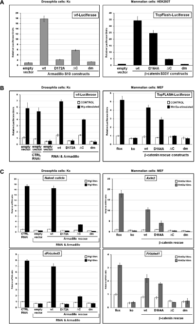
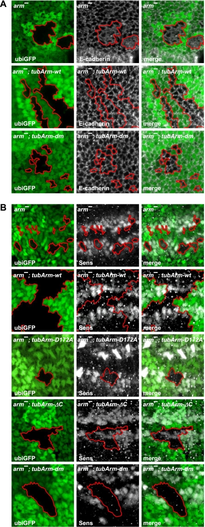
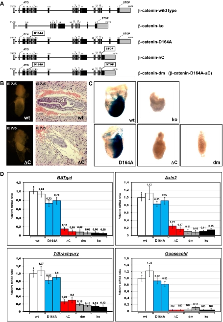
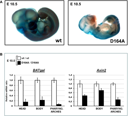
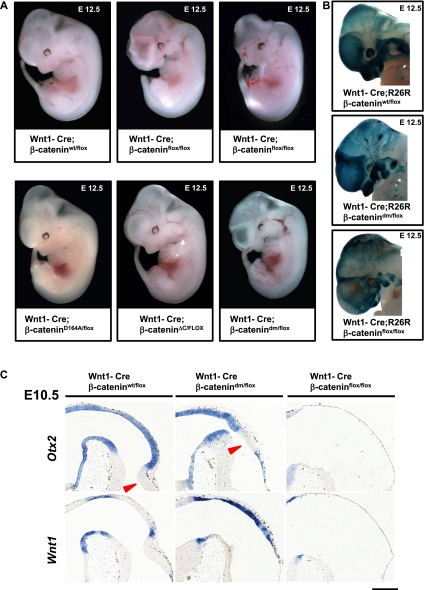
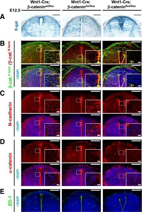
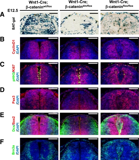
β-Catenin, apart from playing a cell-adhesive role, is a key nuclear effector of Wnt signaling. Based on activity assays in Drosophila, we generated mouse strains where the endogenous β-catenin protein is replaced by mutant forms, which retain the cell adhesion function but lack either or both of the N- and the C-terminal transcriptional outputs. The C-terminal activity is essential for mesoderm formation and proper gastrulation, whereas N-terminal outputs are required later during embryonic development. By combining the double-mutant β-catenin with a conditional null allele and a Wnt1-Cre driver, we probed the role of Wnt/β-catenin signaling in dorsal neural tube development. While loss of β-catenin protein in the neural tube results in severe cell adhesion defects, the morphology of cells and tissues expressing the double-mutant form is normal. Surprisingly, Wnt/β-catenin signaling activity only moderately regulates cell proliferation, but is crucial for maintaining neural progenitor identity and for neuronal differentiation in the dorsal spinal cord. Our model animals thus allow dissecting signaling and structural functions of β-catenin in vivo and provide the first genetic tool to generate cells and tissues that entirely and exclusively lack canonical Wnt pathway activity.


Figures

Figure 1.
Figure 1.
Requirements for N- and C-terminal coactivators for Arm- and β-catenin-mediated transcription. (A–C) The following mutant forms of Arm/β-catenin were tested: wild type (wt), compromised in binding to Lgs/BCL9 (D172A for Arm and D164A for β-catenin), lacking the ability to interact with C-terminal coactivators (ΔC), and the composite of these two individual mutations (dm). (A,B) Luciferase reporter assays in Drosophila Kc and mammalian HEK293T or MEF cells. The activity of Wnt/β-catenin transcription was determined in Kc cells by a wingful-Luciferase (wf-Luciferase) reporter and in mammalian cells by TopFlash. The Y-axes show relative luciferase units normalized to the levels of constantly expressed Renilla. Error bars represent standard deviations. (A) Expression vectors are based on constructs encoding a constitutive active form of Arm (ArmS10) or β-catenin (β-cateninS33Y); the different mutant plasmids were transfected into Kc or HEK293T cells as indicated. Luciferase units shown were normalized relative to those from cells that were transfected in parallel with empty vector (and set to 1). (B) Drosophila Kc cells were treated with dsRNA directed against UTRs of arm mRNA or with control dsRNAi. Cells treated with dsRNAi were transfected with tubulinα1 promoter-driven mutant arm constructs to rescue Arm protein expression. All cells were cultivated with Wg-conditioned medium or control medium. Mammalian MEFs containing a conditional knockout allele of β-catenin (flox) were used for the generation of β-catenin-null MEFs (ko). ko cells were infected with retroviral particles encoding different β-catenin rescue constructs as indicated. Cells were stimulated by Wnt3a-conditioned medium or control medium. Luciferase units shown were normalized relative to those from cells that expressed endogenous Arm or β-catenin in the Wnt-unstimulated situation. (C) Relative expression of the Wg targets Naked cuticle and dFrizzled3 in Drosophila Kc cells (which expressed different mutant forms of Arm instead of endogenous wild-type Arm) and relative expression levels of the Wnt/β-catenin targets Axin2 and Frizzled1 upon Wnt3a treatment in MEFs whose endogenous β-catenin expression was substituted with mutant forms. Cells were generated as in B. mRNA levels were normalized to those of the housekeeping genes SDHA and GAPDH. Y-axes show normalized relative mRNA levels; Wnt-unstimulated cells expressing endogenous Arm and β-catenin, respectively, were set to 1. Error bars show standard deviations.
Figure 2.
Figure 2.
The double-mutant form of Arm restores irregular adherens junctions of arm-null clones, but fails to rescue Wg-mediated transcription in vivo. (A,B) Confocal sections of wing discs are shown that contain arm-null (arm) clones marked by the loss of ubi-GFP expression (borders of clones are highlighted). The particular form of ubiquitously expressed transgenic Arm is indicated in the top corner. Panels to the left show the shape of the clones and the expression of GFP, the middle panels show immunostainings of proteins indicated in the bottom left corner, and the panels to the right show the merged images. (A) Loss of Arm in arm clones results in defective cell shapes and improper adherens junctions as revealed by the adherens junction component E-cadherin. Ubiquitous expression of wild-type Arm fully restores the normal cell adhesion pattern within arm-null clones (arm, tubArm-wt). Expression of Arm lacking the ability to bind both Lgs and C-terminal coactivators sustains normal adhesivity, indistinguishable from the wild-type situation. (B) Arm clones fail to express the Wg target gene senseless (sens). Ubiquitous expression of wild-type Arm (tubArm-wt) fully restores the Sens expression pattern. Arm mutants lacking the ability to bind Lgs (tubArm-D172A) or missing the interaction domain with C-terminal coactivators (tubArm-ΔC) only partially restore Sens expression levels. The double-mutant form of Arm (tubArm-dm) completely lacks the ability to restore Sens expression.
Figure 3.
Figure 3.
Contribution of the C-terminal β-catenin coactivators is essential for gastrulation. (A) Schematic representation of the β-catenin loci of the mutant strains generated. β-Cateninko was generated by crossing a CMV-Cre line with a conditional β-catenin strain (β-cateninflox/flox; i.e., B6.129-Catnbtm2Kem/J); the resulting allele does not contain an in-frame ATG, and exons 2–6, which encode domains essential for binding to E-cadherin and/or TCF/LEF, are eliminated. β-Catenin-D164A harbors a single-amino-acid exchange (D164A) in exon 4, preventing the interaction between the resulting β-catenin and BCL9/BCL9L. In the β-catenin-ΔC locus, a preliminary stop codon is followed by a frameshift introduced into exon 13; moreover, exon 15 was directly fused to exon 13, thus eliminating exon 14. The β-Catenin-dm strain carries both individual mutations. Boxes represent exons, with black indicating coding and gray indicating noncoding; numbers denominate the Arm repeats. (B) Morphology of wild-type (wt) and β-cateninΔC/ΔC (ΔC) embryos at E7.5, including extraembryonic tissues. (Right panels) Embryos dissected from decidual tissues. (Left panels) Sagittal sections of E7.5 embryos within the decidua stained by H&E. (C) Developmental failure during gastrulation caused by C-terminal truncation of β-catenin is associated with absence of TCF/β-catenin-mediated transcription, as monitored by the Wnt-specific reporter BAT-gal. Dissected embryos at E7.5; each individual embryo inherited one allele of the BAT-gal transcriptional reporter and is homozygous for the indicated mutant allele of β-catenin. (ko) Total loss of β-catenin. LacZ expression from the BAT-gal reporter was determined using enzymatic staining based on X-gal (blue). (D) Transcription of Wnt/β-catenin target genes (transgenic reporter BAT-gal and endogenous genes Axin2 and T/Brachyury) is strongly reduced in E6.5 embryos homozygous for the β-catenin allele that prevents the binding of β-catenin to C-terminal transcription coactivators (ΔC). Expression of factors regulating mesoderm formation (T/Brachyury and Goosecoid) is also strongly affected in β-cateninΔC/ΔC embryos. Levels of mRNA were determined by quantitative real-time PCR and normalized to the housekeeping genes SDHA and GAPDH. The levels of a β-cateninwt/wt (wild-type) embryo are set as 1. Embryos from two independent litters were tested for each homozygous β-catenin mutant as indicated. (dm) Double-mutant form; (ko) β-catenin-null embryos. Each embryo carries one allele of the BAT-gal reporter. Error bars show standard deviation. (ND) Nondetectable levels.
Figure 4.
Figure 4.
Developmental defects and reduction in β-catenin-mediated transcription in embryos expressing the D164A mutant form. (A) Wild-type (wt) and homozygous β-cateninD164A/D164A (D164A) embryos at E10.5 carrying one allele of the Wnt/β-catenin transcriptional reporter BAT-gal and stained for enzymatic activity with X-gal (blue). The mutant embryos show severe defects in size and in the development of brain, craniofacial structures, and pharyngeal arches. These defects are associated with reduced BAT-gal activity. Representative embryos are shown. (B) Wnt/β-catenin transcription is reduced in such mutant embryos as determined by quantitative real-time PCR in the head and the pharyngeal arches at E10.0. Expression of the BAT-gal reporter and of the endogenous Wnt/β-catenin target gene Axin2 in the head, pharyngeal arches, and the rest of the body was monitored. Transcript levels are normalized to those of the housekeeping genes SDHA and GAPDH. Levels for wild-type (wt) embryos are set to 1. Error bars show standard deviations. Representative results are shown.
Figure 5.
Figure 5.
Total loss of β-catenin in Wnt1-Cre-expressing tissues leads to more severe phenotypes than blocking the Wnt signaling output of β-catenin. (A) E12.5 embryos expressing Wnt1-Cre, one conditional allele of β-catenin (β-cateninflox), and a second allele of β-catenin as indicated. In Wnt1-Cre; β-cateninflox/flox embryos, Cre activity results in the absence of β-catenin in Wnt1-expressing tissues. Such embryos show strong defects in craniofacial development, midbrain, and hindbrain. The phenotype of Wnt1-Cre; β-catenindm/flox embryos is milder compared with that of Wnt1-Cre; β-cateninflox/flox, especially in the case of telencephalic lobes and midbrain and hindbrain areas. Representative embryos are shown. (B) X-gal staining visualizing Wnt1-Cre-mediated recombination using R26R in the heads of embryos at E12.5. The strong dark-blue signal in forming craniofacial structures (jaw/maxilla and meninges) and pharyngeal arches in wild type represents neural crest derivatives. Similarly strong signals could be partially observed in Wnt1-Cre/R26R; β-catenindm/flox, while in Wnt1-Cre/R26R; β-cateninflox/flox, there are fewer, irregularly scattered X-gal-positive cells. (C) In situ hybridization with early midbrain–hindbrain junction markers on sagittal sections of embryonic heads at E10.5. (Top left panel) In wild-type embryos, the transcription factor Otx2 is expressed in the midbrain with a sharp boundary at the midbrain–hindbrain junction. (Top middle panel) In Wnt1-Cre; β-catenindm/flox, the midbrain–hindbrain junction marked by Otx2 expression is shifted, and the prospective cerebellum is not developed. (Bottom left panel) Wnt1 is expressed in wild type in the posterior midbrain and along the dorsal midline. (Bottom middle panel) Wnt1-Cre; β-catenindm/flox animals show increased expression of Wnt1 along the dorsal midline. (Right panels) In contrast, Wnt1-Cre; β-cateninflox/flox (ko) brains lack expression of both Otx2 (top) and Wnt1 (bottom), indicating absence of any midbrain and cerebellar structures. The arrowheads point to hindbrain structures (cerebellum). Bar, 200 μm.
Figure 6.
Figure 6.
β-Catenin-dm can fully restore the corrupted dorsal spinal cord morphology caused by defective adherens junctions in tissue lacking β-catenin. (A) X-gal staining visualizing Wnt1-Cre-mediated expression of β-galactosidase from R26R in the dorsal spinal cord of E12.5 embryos. Wild-type embryos (Wnt1-Cre/R26R; β-cateninwt/flox) and embryos expressing double-mutant β-catenin (Wnt1-Cre/R26R; β-catenindm/flox) display normal morphology and shape of the dorsal spinal cord. On the other hand, absence of β-catenin (Wnt1-Cre/R26R; β-cateninflox/flox) results in breakage of medial apical contacts and severe morphological defects. (B) Double-mutant β-catenin effectively restores the adhesion defects caused by the loss of β-catenin in Wnt1-Cre-positive tissues. Immunostaining with antibody recognizing either the N terminus or C terminus of β-catenin. The C terminus is missing in the double-mutant form, but protein expression is recognized by the antibody against the N terminus. No β-catenin is detected in the recombination-prone region of Wnt1-Cre; β-cateninflox/flox neural tubes. (C–E) N-cadherin (C), α-catenin (D), and ZO-1 (E) immunostainings for adherens and tight junctions do not show a significant difference between wild-type (Wnt1-Cre; β-cateninwt/flox) and mutant (Wnt1-Cre; β-catenindm/flox) spinal cord. Lack of β-catenin (Wnt1-Cre; β-cateninflox/flox) leads to adhesion defects associated with the breakdown of apical junctional complexes. Bar, 100 μm; bar in inset, 10 μm.
Figure 7.
Figure 7.
Block of Wnt signaling output by double-mutant β-catenin affects cell fate determination in the dorsal spinal cord. (A–F) Immunostaining of transversal sections of dorsal spinal cord isolated from E12.5 embryos. The double-mutant form of β-catenin fully blocks expression of the Wnt reporter BAT-gal (A) and of the endogenous Wnt target gene CyclinD1 (B) in the dorsal spinal cord, where Wnt1-Cre is active. (C) Cells expressing the double-mutant β-catenin are still proliferating, indicated by Ki67 and pHH3 expression. In contrast, cell proliferation was strongly reduced in Wnt1-Cre; β-cateninflox/flox animals. The spinal cord of Wnt1-Cre; β-catenindm/flox animals is characterized by strongly reduced expression of Pax3 (D) and Sox2 (E), indicating a loss of undifferentiated precursor cells. Neuronal differentiation in the dorsal neural tube of these animals is affected as indicated by the absence of early neuronal markers Dcx (E) and Brn3 (F). Bar, 100 μm.

Comment in

References

    1. Arnold SJ, Robertson EJ 2009. Making a commitment: Cell lineage allocation and axis patterning in the early mouse embryo. Nat Rev Mol Cell Biol 10: 91–103 - PubMed
    1. Arnold SJ, Stappert J, Bauer A, Kispert A, Herrmann BG, Kemler R 2000. Brachyury is a target gene of the Wnt/β-catenin signaling pathway. Mech Dev 91: 249–258 - PubMed
    1. Basler K, Struhl G 1994. Compartment boundaries and the control of Drosophila limb pattern by hedgehog protein. Nature 368: 208–214 - PubMed
    1. Bischof J, Maeda RK, Hediger M, Karch F, Basler K 2007. An optimized transgenesis system for Drosophila using germ-line-specific ΦC31 integrases. Proc Natl Acad Sci 104: 3312–3317 - PMC - PubMed
    1. Brault V, Moore R, Kutsch S, Ishibashi M, Rowitch DH, McMahon AP, Sommer L, Boussadia O, Kemler R 2001. Inactivation of the β-catenin gene by Wnt1-Cre-mediated deletion results in dramatic brain malformation and failure of craniofacial development. Development 128: 1253–1264 - PubMed

Publication types