. 2012 Jan 10;109(2):621-6.
doi: 10.1073/pnas.1109237109. Epub 2011 Dec 21.

A role for neuronal cAMP responsive-element binding (CREB)-1 in brain responses to calorie restriction

Affiliations

A role for neuronal cAMP responsive-element binding (CREB)-1 in brain responses to calorie restriction

Salvatore Fusco et al. Proc Natl Acad Sci U S A. .

Abstract

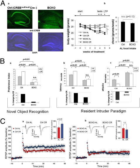
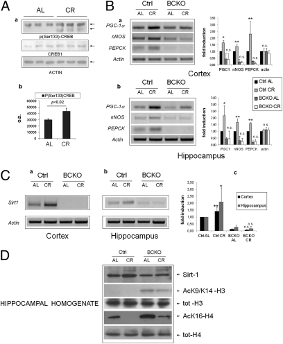
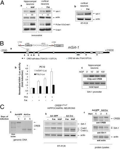
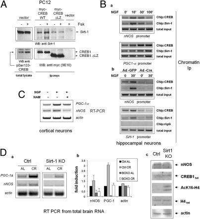
Calorie restriction delays brain senescence and prevents neurodegeneration, but critical regulators of these beneficial responses other than the NAD(+)-dependent histone deacetylase Sirtuin-1 (Sirt-1) are unknown. We report that effects of calorie restriction on neuronal plasticity, memory and social behavior are abolished in mice lacking cAMP responsive-element binding (CREB)-1 in the forebrain. Moreover, CREB deficiency drastically reduces the expression of Sirt-1 and the induction of genes relevant to neuronal metabolism and survival in the cortex and hippocampus of dietary-restricted animals. Biochemical studies reveal a complex interplay between CREB and Sirt-1: CREB directly regulates the transcription of the sirtuin in neuronal cells by binding to Sirt-1 chromatin; Sirt-1, in turn, is recruited by CREB to DNA and promotes CREB-dependent expression of target gene peroxisome proliferator-activated receptor-γ coactivator-1α and neuronal NO Synthase. Accordingly, expression of these CREB targets is markedly reduced in the brain of Sirt KO mice that are, like CREB-deficient mice, poorly responsive to calorie restriction. Thus, the above circuitry, modulated by nutrient availability, links energy metabolism with neurotrophin signaling, participates in brain adaptation to nutrient restriction, and is potentially relevant to accelerated brain aging by overnutrition and diabetes.


Conflict of interest statement

The authors declare no conflict of interest.

Figures

Fig. 1.
Fig. 1.
Impaired brain response to calorie restriction in mice lacking neuronal CREB. (Aa) Immunofluorescence analysis reveals a near complete deletion of CREB1 in the cortex and hippocampus of 6-mo-old BCKO mice. Nuclear staining with DAPI confirms normal cellularity in the same areas. (b) Weight loss under calorie restriction (Left) and food consumption ad libitum (Right) in control (Ctrl) and BCKO mice (n = 5–6 per group, one of two independent experiments); (Left) **P < 0.01; *P < 0.05; n.s. nonsignificant by two-way ANOVA (4 wk time-point); (Right) P by two-tailed t test. (B) Cognitive and behavioral effects of CR. (a) (Upper) Preference toward the novel object in a novel object recognition paradigm. Values (seconds) are mean ± SEM. (Lower) Percent change by CR. (b and c) (Upper) Latency of the first attack (Left, values in seconds, mean ± SEM) and number of attacks in 10 min (Right, rank in an ordinal scale ± SEM) were scored in a resident-intruder paradigm. (Lower) Percentage changes of the corresponding parameter by calorie restriction in control and BCKO mice datasets (n = 6 animals per group) were analyzed by two-way ANOVA; P values are indicated. Experiments were performed twice with similar results. (C) Time course of Schaffer collateral-CA1 LTP induced by tetanic stimulation in control (a) and BCKO (b) mice. Values are percentages of baseline fEPSP amplitude (100%). (Insets) Representative fEPSPs at baseline (dashed line) and during the last 10 min of LTP recording (solid line). Traces are averages of 10 consecutive responses at the time points selected. Bar graphs compare average LTP magnitudes observed during the last 10 min of recording (percentage of baseline fEPSP amplitude). (Student t test, *P < 0.05).
Fig. 2.
Fig. 2.
Brain CREB is activated by calorie restriction and increases the expression of Sirt-1. (Aa) Western blot analysis of hippocampal homogenates from individual mice revealing increased phosphorylation of CREB on Ser-133 by calorie restriction. (b) Band densitometry normalized to actin; statistics by two-tailed t test. (B) RT-PCR analysis of three canonical CREB target mRNAs in the cortex (a) and hippocampus (b) of control and BCKO mice after 4 wk of AL or CR feeding. Actin was used as loading control. Histograms report fold-induction values compared with Ctrl AL (mean ± SD of three to five mice). *P < 0.05; **P < 0.01; n.s., nonsignificant by two-way ANOVA (CR vs. AL). (C) RT-PCR analysis of Sirt-1 mRNA expression and up-regulation by CR in the cortex (a) and hippocampus (b) of control and BCKO mice. Histogram in c displays fold-induction values relative to Ctrl AL (mean ± SD of three to five mice). Statistics as in B. (D) Western blot analysis of hippocampal homogenates showing impaired up-regulation of Sirt-1 by CR and increased acetylation of histones H3 and H4 at Sirt-1-sensitive sites in BCKO mice. Anti-total H3 and H4 histones confirms equal protein input throughout the lanes. Each lane is the pool of hippocampi from two different mice.
Fig. 3.
Fig. 3.
Transcriptional regulation of Sirt-1 by CREB in neuronal cells. (Aa) Immunoblot analysis of whole-cell lysates from primary neurons (hippocampal or cortical) exposed to CREB-activating stimuli. NGF, 50 ng/mL; Fsk, 10 μM. Relevant bands are indicated by arrows. Relative densitometric values for the Sirt-1 band are indicated. CREB phosphorylation and Sirt-1 expression were assayed at different times (30 min and 16 h, respectively). (b) RT PCR analysis of Sirt-1 mRNA in cortical neurons treated with Fsk for 6 h. Actin was amplified as an internal loading control. Panels representative of several independent experiments. (Ba) Scheme displaying several putative CRE elements within the mouse Sirt-1 gene (MGSCv37 C57BL/6J, locus NC_000076). The segment inserted in the Sirt-1-Luc reporter gene, and primers used in ChIP studies (red arrows) are indicated. Two CRE half-sites internal to the segment are also highlighted; numbers are positions relative to the annotated TSS. Exons refer to transcript variant 1. The exon 2 box is shaded because this exon is absent in transcript variants 2 and 3. (b) Luciferase reporter assay confirming responsiveness of the 1738–2180 genomic fragment to Fsk and PKA in PC12 cells. Bars are fold-induction ± SD of triplicate samples; picture representative of two independent experiments. (c) ChIP assay showing NGF-induced binding of CREB to the 1824–2090 Sirt-1 region in hippocampal neurons. Minutes of stimulation are indicated. Sirt-1 promoter was amplified from the total chromatin input as quantitative control (Lower). (Ca) Deletion of CREB exon 10 in cultured CREBloxP/loxP hippocampal neurons 2 or 5 d after adenoviral delivery of Cre recombinase (Ad-Cre). Genomic DNA was amplified with two primers external to the recombination sites. Bands corresponding to undeleted (Upper) and deleted (Lower) alleles are indicated by arrows. (b) RT-PCR analysis revealing defective induction of Sirt-1 mRNA by NGF and Fsk (6 h) in CREB-deleted hippocampal neurons. Actin was amplified as loading control. Bands corresponding to deleted (ΔCREB) and residual undeleted CREB mRNA are indicated by arrows. Picture is representative of several independent experiments. (c) Western blot analysis of whole cell lysates from mock (Ad-GFP) and Cre-infected cells indicating reduced expression of Sirt-1 in the latter cell population. Actin band confirms equal protein loading.
Fig. 4.
Fig. 4.
Sirt-1 promotes CREB-dependent gene expression. (A) Forskolin-inducible physical association of CREB with Sirt-1 in PC12 cells. mycCREB or mycCREB-ΔLZ were transfected in PC12 cells and immunoprecipitated from protein lysates untreated or stimulated with Fsk for 30 min; the presence of Sirt-1 in immunocomplexes was verified by immunoblotting (Upper); anti-Ser133 CREB and anti-myc immunoblotting were used to confirm CREB phosphorylation by Fsk and assess the expression level of transfected CREB isoforms, respectively (Lower). (Ba) ChIP assays showing parallel interaction of CREB and Sirt-1 with the CRE-containing promoter regions of nNOS and PGC-1α in hippocampal neurons treated with NGF. Promoter amplification from total chromatin input is also reported as control. (b) CREB mediates Sirt-1 interaction with CRE-containing promoter regions. NGF-inducible binding of Sirt-1 to nNOS (Upper) and Sirt-1 promoter (Lower) are drastically reduced in hippocampal neurons lacking CREB. Binding of CREB to the same promoter regions confirms severe reduction of CREB binding in Cre-infected cells. rIgG (rabbit IgG) is a negative control for ChIP. Total chromatin input was equal throughout the lanes. (C) RT-PCR analysis showing reduced induction of nNOS and PGC-1α mRNA by NGF in cortical neurons treated with the Sirt-1 inhibitor Nicotinamide (NAM). (D) (a and b) Representative RT-PCR analysis of PGC-1α and nNOS mRNA expression in whole brains from WT and Sirt-1-deficient mice under both AL and CR (6 mo) feeding. Each lane represents a pool of two mice. Actin was used as loading control. (c) Western blot analysis of whole brain protein homogenates from WT and Sirt-1 KO mice (fed AL) indicating reduced expression of nNOS but normal levels of immunoreactive CREB and total histone H4. Anti-AcH4K16 and antiactin immunostaining confirm, respectively, reduced deacetylase activity in the SirtKO sample and equal protein loading in the two lanes.

References

    1. Fontana L, Partridge L, Longo VD. Extending healthy life span—From yeast to humans. Science. 2010;328:321–326. - PMC - PubMed
    1. Maalouf M, Rho JM, Mattson MP. The neuroprotective properties of calorie restriction, the ketogenic diet, and ketone bodies. Brain Res Brain Res Rev. 2009;59:293–315. - PMC - PubMed
    1. Takeda S, et al. Diabetes-accelerated memory dysfunction via cerebrovascular inflammation and Abeta deposition in an Alzheimer mouse model with diabetes. Proc Natl Acad Sci USA. 2010;107:7036–7041. - PMC - PubMed
    1. de la Monte SM. Insulin resistance and Alzheimer's disease. BMB Rep. 2009;42:475–481. - PMC - PubMed
    1. Haigis MC, Guarente LP. Mammalian sirtuins—Emerging roles in physiology, aging, and calorie restriction. Genes Dev. 2006;20:2913–2921. - PubMed

Publication types

MeSH terms

LinkOut - more resources