. 2012 Feb;139(3):514-24.
doi: 10.1242/dev.071746. Epub 2011 Dec 21.

Wnt/β-catenin signaling directly regulates Foxj1 expression and ciliogenesis in zebrafish Kupffer's vesicle

Affiliations

Wnt/β-catenin signaling directly regulates Foxj1 expression and ciliogenesis in zebrafish Kupffer's vesicle

Alissa Caron et al. Development. 2012 Feb.

Abstract

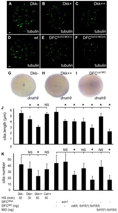
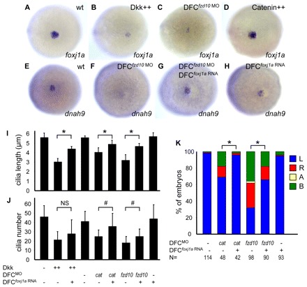
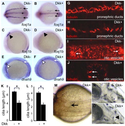
Cilia are essential for normal development. The composition and assembly of cilia has been well characterized, but the signaling and transcriptional pathways that govern ciliogenesis remain poorly studied. Here, we report that Wnt/β-catenin signaling directly regulates ciliogenic transcription factor foxj1a expression and ciliogenesis in zebrafish Kupffer's vesicle (KV). We show that Wnt signaling acts temporally and KV cell-autonomously to control left-right (LR) axis determination and ciliogenesis. Specifically, reduction of Wnt signaling leads to a disruption of LR patterning, shorter and fewer cilia, a loss of cilia motility and a downregulation of foxj1a expression. However, these phenotypes can be rescued by KV-targeted overexpression of foxj1a. In comparison to the FGF pathway that has been previously implicated in the control of ciliogenesis, our epistatic studies suggest a more downstream function of Wnt signaling in the regulation of foxj1a expression and ciliogenesis in KV. Importantly, enhancer analysis reveals that KV-specific expression of foxj1a requires the presence of putative Lef1/Tcf binding sites, indicating that Wnt signaling activates foxj1a transcription directly. We also find that impaired Wnt signaling leads to kidney cysts and otolith disorganization, which can be attributed to a loss of foxj1 expression and disrupted ciliogenesis in the developing pronephric ducts and otic vesicles. Together, our data reveal a novel role of Wnt/β-catenin signaling upstream of ciliogenesis, which might be a general developmental mechanism beyond KV. Moreover, our results also prompt a hypothesis that certain developmental effects of the Wnt/β-catenin pathway are due to the activation of Foxj1 and cilia formation.


Figures

Fig. 1.
Fig. 1.
Wnt/β-catenin signaling regulates LR asymmetry KV cell-autonomously. (A-C) fzd10 expression pattern. Shown are a lateral view of 80% epiboly (A), and lateral (B) and dorsal (C) views of the tail bud region of 10-somite staged zebrafish embryos with anterior to the left. Arrow, DFCs; arrowhead, KV. (D) Western blot showing diminished Fzd10 level in fzd10 morphants. Actin was used as a loading control. (E-H) fzd10 transduces Wnt/β-catenin signaling. DFC-specific injection of fzd10 MO depleted sp5l expression from DFCs (F). Knockdown of Fzd10 resulted in downregulation of axin2 expression (H). Shown are lateral views of 80% epiboly (E,F) and 10-somite (G,H) staged embryos. Arrow indicates DFCs. (I) Representative images of spaw expression (bracket) in DFCfzd10 MO embryos at the 21-somite stage. (J) DFC/KV-specific reduction of Wnt/β-catenin signaling randomizes spaw expression. Percentages of spaw expression were determined in 21-somite staged embryos. MO (ng), amount of MO used; N, number of embryos examined. L, left-side; R, right-side; A, absence; B, bilateral. *P<0.01 compared with corresponding modulations in the yolk only or with uninjected controls.
Fig. 2.
Fig. 2.
Reduction of Wnt signaling results in shorter and fewer cilia in KV. (A-F) Dose-dependent effect of Wnt signaling on cilia length and number. (A-C) Tg(hsp:dkk1-GFP) fish were bred with wild-type fish. Their progenies were heat shocked at 60% epiboly for 30 minutes (B) or 60 minutes (C) to induce Dkk1 expression. Moderate induction of Dkk1 (Dkk+) resulted in shorter cilia (B) whereas higher level of Dkk1 induction (Dkk++) led to shorter and fewer cilia (C). (D-F) DFC-targeted injection of 1 ng of fzd10 MO reduced cilia length, but not number (E); 4 ng of MO reduced both cilia length and number (F). Cilia were visualized by anti-acetylated tubulin antibody staining of 10-somite staged embryos. Scale bars: 5 μm. (G-I) Wnt signaling is essential for dnah9 expression in DFCs. Dkk1 induction (H) or DFC-targeted injection of β-catenin1 MO (I) downregulated dnah9 expression. Shown are ventral views of 95% epiboly staged embryos. (J,K) Quantification of cilia length (J) and number (K) in 10-somite staged embryos. Approximately 12-16 embryos were analyzed for each group. Tg(hsp:β-catenin-GFP) fish were bred with wild-type fish. Their progenies were heat shocked at 60% epiboly for 60 minutes to induce β-catenin1 (Cat++) expression. Data are represented as mean±s.d. *P<0.01. NS, not statistically significant.
Fig. 3.
Fig. 3.
Wnt signaling regulates KV ciliogenesis through modulation of foxj1a expression. (A-D) Wnt signaling is required for foxj1a expression in DFCs. foxj1a expression in DFCs was severely downregulated in Dkk1-expressing zebrafish embryos (B; 37/38) and embryos injected with fzd10 MO into DFCs (C; 24/27) compared with wild-type controls (A). Induction of β-catenin1 did not appear to affect foxj1a expression (D). (E-H) Ectopic expression of foxj1a restores dnah9 levels in embryos with impaired Wnt activity. Downregulated dnah9 expression in DFCfzd10 MO embryos (F; 16/18) was enhanced by DFC-targeted foxj1a overexpression (G; 15/19). Overexpression of foxj1a RNA alone did not significantly alter dnah9 level (H). Shown are ventral views of 90-95% epiboly staged embryos. (I,J) Ectopic expression of foxj1a partially rescues KV cilia length (I) and, to a lesser extent, cilia number (J). Approximately 10-18 embryos for each group were analyzed at the 10-somite stage. Data are represented as mean±s.d. *P<0.01; #P<0.05. NS, not significant (P>0.05). Tg(hsp:dkk1-GFP) and Tg(hsp:β-catenin-GFP) embryos were heat shocked at 50% epiboly for 60 minutes. (K) Ectopic expression of foxj1a partially restores left-sided spaw expression in embryos with impaired Wnt activity. Percentages of spaw expression were determined in 18-to 21-somite staged embryos. N, number of embryos examined. L, left-side; R, right-side; A, absence; B, bilateral.
Fig. 4.
Fig. 4.
Wnt signaling functions downstream of the FGF pathway in regulation of foxj1a expression and cilia formation. (A-D) Ectopic expression of Wnt ligands restores foxj1a levels in embryos with reduced FGF signaling. DFCfgfr1 MO embryos exhibited downregulated foxj1a expression in DFCs (B; 17/18) compared with controls (A). Co-injection of wnt8a RNA with fgfr1 MO restored foxj1a expression (C; 16/19). Ectopic expression of wnt8a RNA alone had no apparent effect on foxj1a expression (D). (E-H) Ectopic expression of FGF ligands fails to restore foxj1a expression in embryos with reduced Wnt signaling. Downregulated foxj1a expression in DFCfzd10 MO embryos (F) was not restored by co-injection of fgf8a RNA (G). Ectopic expression of fgf8a RNA alone had no apparent effect on foxj1a expression (H). Shown are ventral views of 90-95% epiboly staged embryos. (I,J) Ectopic expression of Wnt ligands partially rescues cilia length defects in embryos with reduced FGF signaling. Shown are quantification of cilia length (I) and cilia number (J). Approximately 12-20 embryos were analyzed for each group. Data are represented as mean±s.d. *P<0.01. NS, not significant (P>0.05).
Fig. 5.
Fig. 5.
Wnt signaling directly regulates foxj1a transcription. (A) Schematic of foxj1a enhancers. Sequence of foxj1a that is located approximately –6.2 kb to –4.6 kb upstream of the ATG start codon was used to generate a series of report constructs. Blue bar, conserved sequence between zebrafish and tetradon. Hatched bar, predicted first non-coding exon. Red line, putative Lef1/Tcf binding site. These fragments were inserted in front of the gfp sequence in a tol2 vector, and the resulting plasmids were co-injected with tol2 transposase RNA into embryos. Fluorescent GFP signals in KV were scored. (B,C) A 0.6 kb enhancer sequence contains Wnt-responsive cis-acting elements. Transgenic Tg(0.6foxj1a:gfp) embryos showed reporter gfp expression in DFCs (B; 12/12). DFC-targeted injection of fzd10 MO abolished gfp expression in DFCs (C; 14/15). Shown is in situ hybridization using gfp as a probe in a ventral view of 95% epiboly staged embryos. Dashed circle indicates DFC region. (D-I) Putative Lef1/Tcf binding sites are required for foxj1a expression in KV. GFP reporter expression in stable transgenic Tg(0.6foxj1a:gfp) embryos recapitulated foxj1a expression pattern in KV (D,E), PDs (E,F) and FP (E,F). After all three putative Lef1/Tcf binding sites in the 0.6 kb enhancer were deleted using a site-directed mutagenesis kit, stable transgenic Tg(0.6Δfoxj1a:gfp) embryos lacked GFP expression in KV (G,H) and PDs (H,I) but maintained GFP expression in FP (H,I). Arrow indicates KV, open arrowhead PDs and filled arrowhead FP. Shown are embryos at 10-12 somites (D,G), 16 somites (E,H) and 30 hpf (F,I).
Fig. 6.
Fig. 6.
Dkk1 induction impairs ciliogenesis in PD and OV. (A-D) Induction of Dkk1 downregulates foxj1 expression in PDs and OVs. foxj1a level in PDs was downregulated in transgenic Dkk1 embryos (B; 8/8) versus non-transgenic siblings (A). foxj1a expression in FP was not affected (B). foxj1b expression in the otic placode was downregulated in transgenic Dkk1 embryos (D; 8/8) versus non-transgenic siblings (C). Arrow indicates PDs, asterisk FP, and arrowhead the otic placode. (E,F) Induction of Dkk1 suppressed dnah9 expression in PDs and OVs (F; 11/11) compared with controls (E). White arrow represents PDs, and white arrowhead the otic placode. Shown are dorsal views (A,B) and lateral views (C-F) of 10-somite staged embryos with anterior to the left. (G-J) Induction of Dkk1 results in shorter and fewer cilia in PDs and OVs. Cilia were visualized by anti-tubulin antibody staining of 26-somite staged embryos. White arrow indicates tethering cilia in OVs. (K,L) Quantification of cilia length in PDs (K) and OVs (tethering cilia) (L). Data are represented as mean±s.d. Approximately 9-18 embryos were used for each group. *P<0.01. (M-O) Induction of Dkk1 results in kidney cysts and otolith malformation. Cystic distension of PD (arrow in M; 45/73) was observed at 24 hpf. One otolith (arrowhead in O; 89/95) was seen at 2 dpf. Tg(hsp:dkk1-GFP) embryos were heat activated at 60% epiboly.

References

    1. Aamar E., Dawid I. B. (2008). Isolation and expression analysis of foxj1 and foxj1.2 in zebrafish embryos. Int. J. Dev. Biol. 52, 985–991 - PMC - PubMed
    1. Afzelius B. A. (1976). A human syndrome caused by immotile cilia. Science 193, 317–319 - PubMed
    1. Alexander J., Rothenberg M., Henry G. L., Stainier D. Y. (1999). Casanova plays an early and essential role in endoderm formation in zebrafish. Dev. Biol. 215, 343–357 - PubMed
    1. Amack J. D., Yost H. J. (2004). The T box transcription factor no tail in ciliated cells controls zebrafish left-right asymmetry. Curr. Biol. 14, 685–690 - PubMed
    1. Badano J. L., Mitsuma N., Beales P. L., Katsanis N. (2006). The ciliopathies: an emerging class of human genetic disorders. Annu. Rev. Genomics Hum. Genet. 7, 125–148 - PubMed

Publication types

MeSH terms

LinkOut - more resources