. 2012 Feb;23(4):716-28.
doi: 10.1091/mbc.E11-06-0530. Epub 2011 Dec 21.

LAS1L interacts with the mammalian Rix1 complex to regulate ribosome biogenesis

Affiliations

LAS1L interacts with the mammalian Rix1 complex to regulate ribosome biogenesis

Christopher D Castle et al. Mol Biol Cell. 2012 Feb.

Abstract

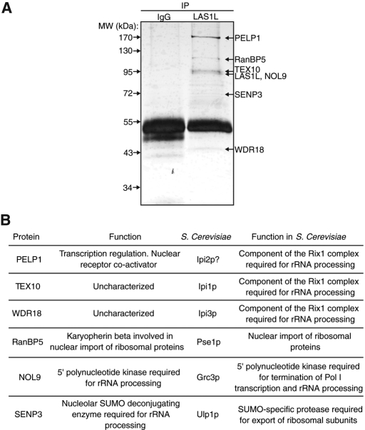
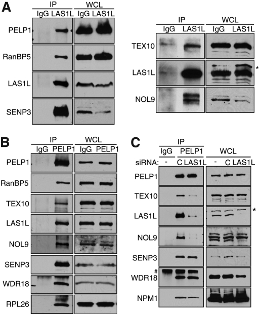
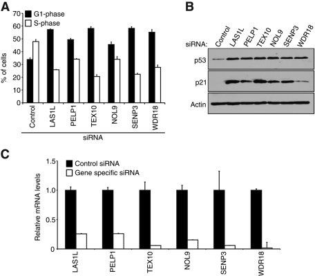
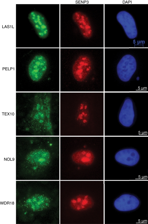
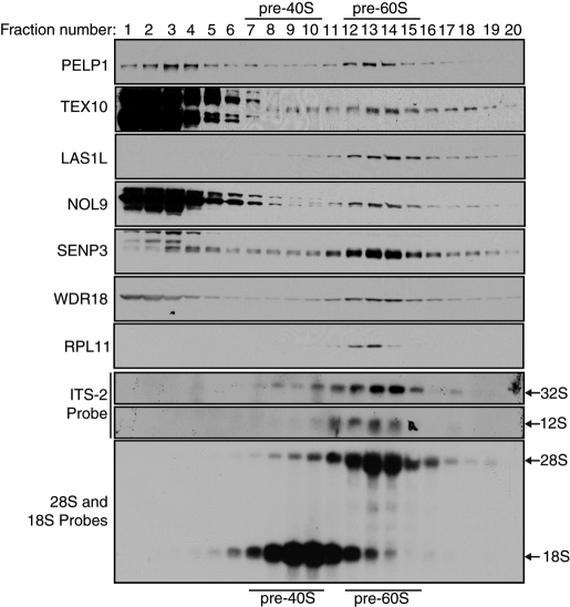
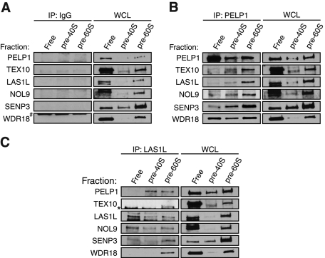
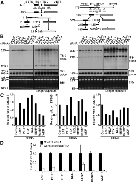
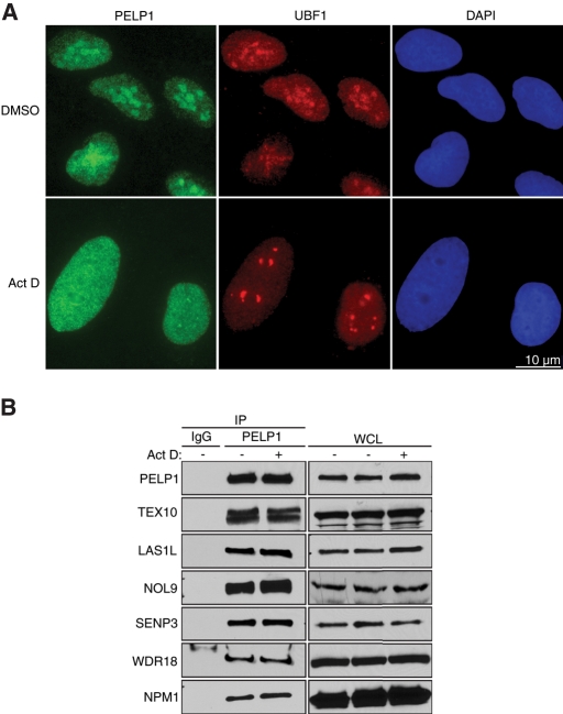
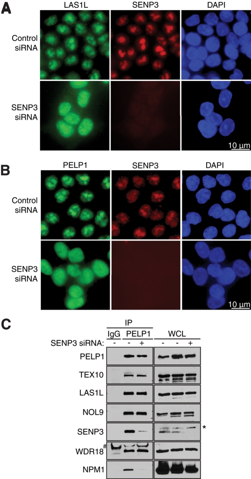
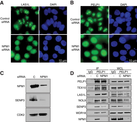
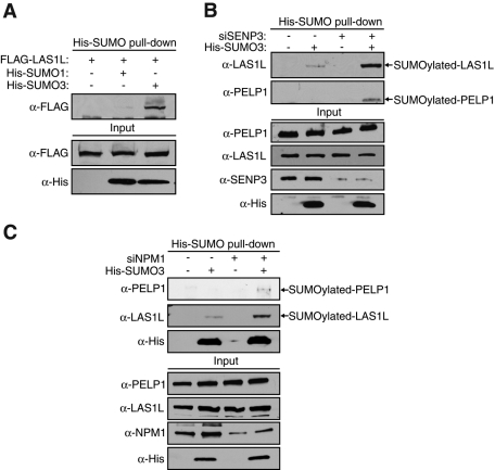
The coordination of RNA polymerase I transcription with pre-rRNA processing, preribosomal particle assembly, and nuclear export is a finely tuned process requiring the concerted actions of a number of accessory factors. However, the exact functions of some of these proteins and how they assemble in subcomplexes remain poorly defined. LAS1L was first described as a nucleolar protein required for maturation of the 60S preribosomal subunit. In this paper, we demonstrate that LAS1L interacts with PELP1, TEX10, and WDR18, the mammalian homologues of the budding yeast Rix1 complex, along with NOL9 and SENP3, to form a novel nucleolar complex that cofractionates with the 60S preribosomal subunit. Depletion of LAS1L-associated proteins results in a p53-dependent G1 arrest and leads to defects in processing of the pre-rRNA internal transcribed spacer 2 region. We further show that the nucleolar localization of this complex requires active RNA polymerase I transcription and the small ubiquitin-like modifier-specific protease SENP3. Taken together, our data identify a novel mammalian complex required for 60S ribosomal subunit synthesis, providing further insight into the intricate, yet poorly described, process of ribosome biogenesis in higher eukaryotes.


Figures

FIGURE 1:
FIGURE 1:
Isolation of LAS1L-associated proteins. (A) LAS1L was affinity-purified from HEK 293T cell lysate using an anti-LAS1L antibody coupled to protein G Sepharose beads. Normal rabbit IgG was used as negative control. LAS1L-associated complexes were separated on SDS–PAGE and silver-stained and bands present only in the LAS1L IP lane were cut out, digested with trypsin, and analyzed by mass spectrometry. Identified proteins are marked with arrows. (B) The proteins identified in (A) and their corresponding S. cerevisiae putative homologues (Rout et al., 1997; Jakel and Gorlich, 1998; Bassler et al., 2001; Vadlamudi et al., 2001, 2004; Peng et al., 2003; Galani et al., 2004; Krogan et al., 2004; Gong and Yeh, 2006; Panse et al., 2006; Nair et al., 2007; Haindl et al., 2008; Yun et al., 2008; Braglia et al., 2010; Chou et al., 2010; Heindl and Martinez, 2010; Finkbeiner et al., 2011).
FIGURE 2:
FIGURE 2:
LAS1L associates with PELP1, TEX10, WDR18, RanBP5, NOL9, and SENP3. (A) LAS1L was immunoprecipitated (IP) with an anti-LAS1L–specific antibody from HEK 293T cell lysates. Associated proteins were separated on SDS–PAGE and analyzed by Western blotting with specific antibodies (indicated on the left). Normal rabbit IgG was used as negative control. WCL, whole-cell lysate; *, presence of an unspecific band. (B) PELP1 was immunoprecipitated (IP) with an anti-PELP1 antibody from HEK 293T cell lysates. Associated proteins were separated on SDS–PAGE and analyzed by Western blotting with specific antibodies (indicated on the left). Normal rabbit IgG was used as negative control. WCL, whole-cell lysate. (C) HEK 293T cells were transfected with nontargeting control (represented by the letter “C”) or LAS1L siRNA for 48 h. Cells were lysed and immunoprecipitated with an anti-PELP1 antibody. Proteins associating with PELP1 in the absence of LAS1L were separated on SDS–PAGE and analyzed by Western blotting with specific antibodies (indicated on the left). Normal rabbit IgG was used as negative control. WCL, whole-cell lysate; *, presence of an unspecific band; #, an IgG band.
FIGURE 3:
FIGURE 3:
Depletion of LAS1L-interacting proteins induces a p53-dependent G1 cell cycle arrest. (A) Cell cycle profiles of HCT116 cells transfected with control, LAS1L, PELP1, TEX10, NOL9, SENP3, and WDR18 siRNA. Seventy-two hours after transfection the cells were pulse-labeled with BrdU for 30 min, stained with propidium iodide, and analyzed by flow cytometry to determine the percentage of cells in G1 and S phase. Error bars indicate SD from triplicate experiments. (B) Western blot analysis of the siRNA-transfected cells from (A) with specific antibodies against p53, p21, and β-actin. (C) Total RNA was extracted from the siRNA-transfected cells from panel (A), and knockdowns were confirmed by qRT-PCR with gene-specific primers. Relative mRNA levels for each gene-specific primer were normalized to β-actin. Error bars indicate SD from triplicate experiments.
FIGURE 4:
FIGURE 4:
LAS1L-associated proteins localize to the nucleolus. Immunofluorescence analysis of U2OS cells with complex protein-specific antibodies. Cells were preextracted with 0.1% Triton, fixed, and immunostained with anti-LAS1L, PELP1, TEX10, NOL9, and WDR18 antibodies (green). Colocalization with SENP3 was confirmed using an anti-SENP3 antibody (red). DNA was visualized by staining with Hoechst 33342 (blue). Scale is representative of all three panels.
FIGURE 5:
FIGURE 5:
LAS1L-interacting proteins cosediment with pre-60S ribosomal particles. Nuclear extracts from HCT116 cells were fractionated by centrifugation on a 10–30% sucrose gradient. Fractions were collected, and the optical density was measured at 260 nm (A260). The positions of the pre-40S and pre-60S native subunits are indicated. Presence of the LAS1L complex proteins in each fraction were confirmed by Western blotting using specific antibodies (indicated on the left). Total RNA from each fraction was extracted and separated on a 1.2% formaldehyde gel and analyzed by Northern blotting with ITS-2, 28S, and 18S probes, as indicated. Arrows indicate the positions of the 32S, 28S, 18S, and 12S rRNAs.
FIGURE 6:
FIGURE 6:
LAS1L and NOL9 interact with the mammalian Rix1 complex on pre-60S ribosomal particles. Nuclear extracts from HCT116 cells were fractionated by centrifugation on a 10–30% sucrose gradient. Fractions were collected, and the optical density was measured at 260 nm (A260). Based on the A260 profile, fractions corresponding to free nuclear proteins (1, 2, and 3), pre-40S ribosomal particles (8, 9, and 10), and pre-60S ribosomal particles (12, 13, and 14) were then combined and immunoprecipitated (IP) with rabbit IgG (A) as a negative control or with PELP1 (B) or LAS1L (C) antibodies. Associated proteins were separated on SDS–PAGE and analyzed by Western blotting with specific antibodies (indicated on the left). WCL, whole-cell lysate; *, presence of an unspecific band; #, an IgG band.
FIGURE 7:
FIGURE 7:
LAS1L-associated proteins are required for proper processing of ITS-2. (A) Schematic representation of the primary 47S rRNA transcript and the two major processing pathways with rRNA intermediates, as indicated (adapted from Hadjiolova et al., 1993). (B) Northern blot analysis of total RNA from HCT116 cells transfected with control, LAS1L, PELP1, TEX10, NOL9, SENP3, RanBP5, and WDR18 siRNA. Seventy-two hours after transfection, equal amounts of total RNA were hybridized with specific probes for ITS-1, ITS-2, 28S, and 18S rRNA intermediates (indicated on the right). The right panel shows longer exposure times for each probe. The positions of the specific probes used for Northern blot analysis are indicated on the schematic in (A). (C) The 32S/28S, 12S/28S, and 30S/28S ratios were determined by quantification of the relative band intensities of the 32S, 30S, and 28S rRNA intermediates in the lower exposures and the 12S rRNA intermediate in the longer exposure from (B). Intensities were normalized to the control siRNA-treated sample. (D) Knockdowns were confirmed by qRT-PCR with gene-specific primers. Relative mRNA levels for each gene-specific primer were normalized to β-actin. Error bars indicate SD from triplicate experiments.
FIGURE 8:
FIGURE 8:
PELP1 requires active Pol I transcription for nucleolar localization. (A) U2OS cells were treated with either DMSO or 20 nM actinomycin D for 2 h. Cells were fixed and immunostained with an anti-PELP1 antibody (green) and an anti-UBF1 antibody (red). DNA was visualized by staining with Hoechst 33342 (blue). Scale is representative of both panels. (B) Cells were treated with DMSO (−) or actinomycin D (+) as in (A), and lysates were immunoprecipitated (IP) with an anti-PELP1 antibody. Associated proteins were separated on SDS–PAGE and analyzed by Western blotting with specific antibodies (indicated on the left). Normal rabbit IgG was used as negative control. WCL, whole-cell lysate.
FIGURE 9:
FIGURE 9:
SENP3 is necessary for LAS1L and PELP1 nucleolar localization. (A) HCT116 cells were transfected with control or SENP3 siRNA for 72 h. Cells were fixed and immunostained with anti-LAS1L (green) and anti-SENP3 (red) antibodies. DNA was visualized by staining with Hoechst 33342 (blue). Scale is representative of all panels. (B) HCT116 cells were transfected with control or SENP3 siRNA for 72 h. Cells were fixed and immunostained with anti-PELP1 (green) and anti-SENP3 (red) antibodies. DNA was visualized by staining with Hoechst 33342 (blue). Scale is representative of all panels. (C) Cells were transfected with a Control (−) or SENP3 (+) siRNA for 72 h. Lysates were then immunoprecipitated with rabbit IgG (as negative control) or PELP1 antibody. Coprecipitating proteins were separated on SDS–PAGE and analyzed by Western blotting using specific antibodies (indicated on the left). WCL, whole-cell lysate; *, presence of an unspecific band; #, an IgG band.
FIGURE 10:
FIGURE 10:
NPM1 is required for LAS1L and PELP1 nucleolar localization. (A) HCT116 cells were transfected with control or NPM1 siRNA for 48 h. Subcellular localization of LAS1L was determined by immunofluorescence analysis using a LAS1L antibody (green). DNA was visualized by staining with Hoechst 33342 (blue). Scale is representative of all panels. (B) HCT116 cells were transfected with control or NPM1 siRNA for 48 h. Subcellular localization of PELP1 was determined by immunofluorescence analysis using a PELP1 antibody (green). DNA was visualized by staining with Hoechst 33342 (blue). Scale is representative of all panels. (C) Knockdowns of NPM1 for (A) and (B) were confirmed by Western blotting using specific antibodies (indicated on the left). An anti-CDK2 antibody was used as loading control. (D) Cells were transfected with a control (“C”) or NPM1 siRNA for 72 h. Lysates were then immunoprecipitated with rabbit IgG (as negative control) or PELP1 antibody. Coprecipitating proteins were separated on SDS–PAGE and analyzed by Western blotting using specific antibodies (indicated on the left). WCL, whole-cell lysate.
FIGURE 11:
FIGURE 11:
LAS1L and PELP1 are modified by SUMO in an SENP3-dependent manner. (A) HEK 293T cells were transfected with FLAG-LAS1L (+) plus either empty vector (−) or plasmids expressing 6xHis-tagged SUMO-1 or SUMO-3 (+). Forty-eight hours after transfection, SUMOylated proteins were pulled down from cell lysates using Ni-NTA agarose beads. Eluates were analyzed on SDS–PAGE and by Western blotting with the indicated antibodies. (B) HEK 293T cells were transfected with either control (−) or SENP3 (+) siRNA. The next day, cells were transfected with empty vector (−) or a 6xHis-tagged SUMO-3 (+) expressing plasmid. Forty-eight hours after transfection, pulldowns and protein analyses were performed as in (A). (C) HEK 293T cells were transfected with either control (−) or NPM1 (+) siRNA. The next day, cells were transfected with empty vector (−) or a plasmid expressing 6xHis-tagged SUMO-3 (+). Forty-eight hours after transfection, pulldowns and protein analyses were performed as in (A).

References

    1. Arabi A, et al. c-Myc associates with ribosomal DNA and activates RNA polymerase I transcription. Nat Cell Biol. 2005;7:303–310. - PubMed
    1. Bassler J, Grandi P, Gadal O, Lessmann T, Petfalski E, Tollervey D, Lechner J, Hurt E. Identification of a 60S preribosomal particle that is closely linked to nuclear export. Mol Cell. 2001;8:517–529. - PubMed
    1. Boisvert FM, van Koningsbruggen S, Navascues J, Lamond AI. The multifunctional nucleolus. Nat Rev Mol Cell Biol. 2007;8:574–585. - PubMed
    1. Braglia P, Heindl K, Schleiffer A, Martinez J, Proudfoot NJ. Role of the RNA/DNA kinase Grc3 in transcription termination by RNA polymerase I. EMBO Rep. 2010;11:758–764. - PMC - PubMed
    1. Budde A, Grummt I. p53 represses ribosomal gene transcription. Oncogene. 1999;18:1119–1124. - PubMed

MeSH terms