. 2012 Feb;18(2):241-52.
doi: 10.1261/rna.030338.111. Epub 2011 Dec 21.

Characterization of the stop codon readthrough signal of Colorado tick fever virus segment 9 RNA

Affiliations

Characterization of the stop codon readthrough signal of Colorado tick fever virus segment 9 RNA

Sawsan Napthine et al. RNA. 2012 Feb.

Abstract

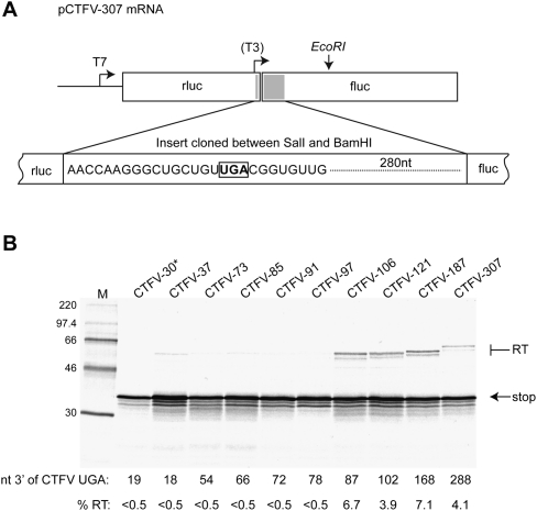
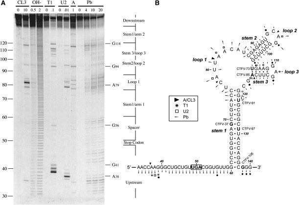
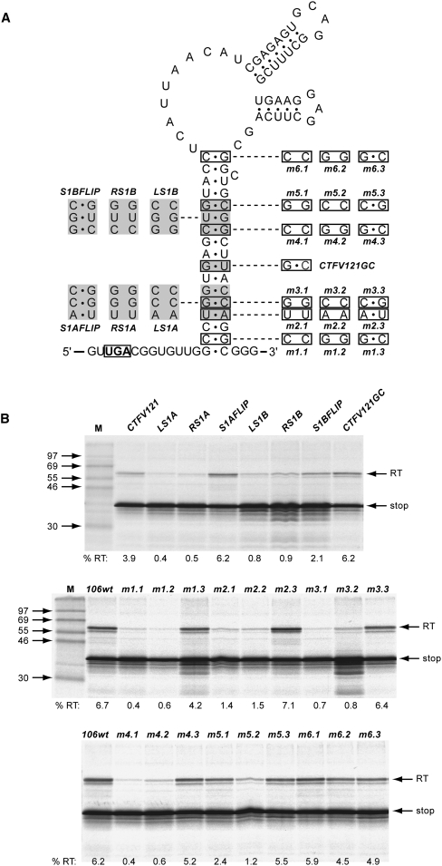
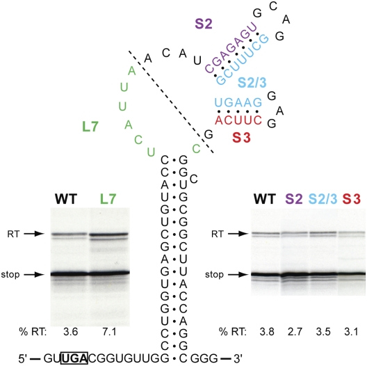
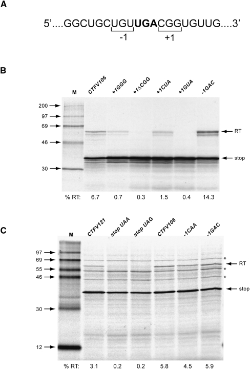
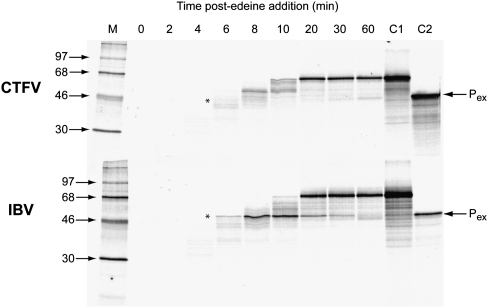
Termination codon readthrough is utilized as a mechanism of expression of a growing number of viral and cellular proteins, but in many cases the mRNA signals that promote readthrough are poorly characterized. Here, we investigated the readthrough signal of Colorado tick fever virus (CTFV) segment 9 RNA (Seg-9). CTFV is the type-species of the genus Coltivirus within the family Reoviridae and is a tick-borne, double-stranded, segmented RNA virus. Seg-9 encodes a 36-kDa protein VP9, and by readthrough of a UGA stop codon, a 65-kDa product, VP9'. Using a reporter system, we defined the minimal sequence requirements for readthrough and confirmed activity in both mammalian and insect cell-free translation systems, and in transfected mammalian cells. Mutational analysis revealed that readthrough was UGA specific, and that the local sequence context around the UGA influenced readthrough efficiency. Readthrough was also dependent upon a stable RNA stem-loop structure beginning eight bases downstream from the UGA codon. Mutational analysis of this stem-loop revealed a requirement for the stem region but not for substructures identified within the loop. Unexpectedly, we were unable to detect a ribosomal pause during translation of the CTFV signal, suggesting that the mechanism of readthrough, at least at this site, is unlikely to be dependent upon RNA secondary-structure induced ribosomal pausing at the recoded stop codon.


Figures

FIGURE 1.
FIGURE 1.
Characterization of the minimal sequence requirements for CTFV readthrough. (A) Schematic of the pCTFV-307 reporter mRNA. A portion of the CTFV Seg-9 sequence (indicated as a gray box) encompassing the VP9 UGA stop codon (16 nt upstream of and 208 nt downstream from the UGA) was cloned into the SalI and BamHI sites of the p2luc reporter plasmid. Transcripts for in vitro translation were generated using T7 RNA polymerase and EcoRI-cut pCTFV-307 and derivatives. The location of the T3 promoter present in the structure mapping construct pCTFV-121-T3 is indicated. (B) Deletion analysis of the CTFV readthrough signal. A series of reporter plasmid variants were prepared with stepwise, in-frame deletions from the 3′ end of the inserted viral sequence. Plasmids were linearized with EcoRI and run-off transcripts translated in Flexi RRL at a final RNA concentration of 50 μg/mL in the presence of [35S]methionine and 140 mM added KCl. The products were resolved by 12% SDS-PAGE and visualized by autoradiography. Molecular size markers were also run on the gel (M). The number of nucleotides 3′ of the CTFV UGA is shown below the gel. Products derived from termination at the CTFV UGA (stop) or following readthrough (RT) are indicated by arrows. The readthrough efficiency of each mRNA is indicated (%RT). The asterisk (CTFV-30) highlights that this mRNA has a shorter stretch upstream of the UGA (11 nt) in comparison to the other mRNAs (16 nt).
FIGURE 2.
FIGURE 2.
Structure probing of the CTFV readthrough signal. (A) RNA derived by transcription of pCTFV-121-T3/BamHI with T3 RNA polymerase was 5′ end-labeled with [γ-33P]ATP and subjected to limited RNase or chemical cleavage using structure-specific probes. Sites of cleavage were identified by comparison with a ladder of bands created by limited alkaline hydrolysis of the RNA (OH-; RNA heated to 100°C for 0.5 or 2 min) and the position of known RNase U2 and T1 cuts, determined empirically. Products were analyzed on a 10% acrylamide/7M urea gel containing formamide. Enzymatic structure probing was with RNases CL3, T1, U2, and A. Uniquely cleaved nucleotides were identified by their absence in untreated control lanes (0). The number of units of enzyme added to each reaction is indicated. Chemical structure probing was with lead acetate (Pb; mM concentration in reaction). (B) The sequence of the probed CTFV RNA and the inferred secondary structure. The sensitivity of bases in the CTFV readthrough region to the various probes is shown for an mfold prediction. The first base of the transcript is numbered 1. The reactivies of the T1 (asterisk), U2 (open square), A and CL3 (black triangle) probes are marked. Lead cleavages are indicated by thin arrows. The size of the symbols is approximately proportional to the intensity of cleavage at that site. Also indicated is the location of the 3′ edge of the truncated versions of CTFV-307 (labeled as in Fig. 1), with the last viral base in each truncation emboldened.
FIGURE 3.
FIGURE 3.
The role of the CTFV stimulatory RNA stem 1 region in readthrough. (A) Complementary and compensatory changes to base pairs within stem 1 were prepared, targeting either blocks of three consecutive base pairs (based on CTFV-121 and shown to the left of the main stem) or single base pairs (based on CTFV-106 and shown to the right of the main stem). In CTFV121GC, the central GU pair was changed to GC. This mutation was also present in RS1A and S1AFLIP. (B) Plasmids containing the mutations detailed in A were linearized, transcribed, translated, and analyzed according to the legend of Figure 1.
FIGURE 4.
FIGURE 4.
Investigating the role of the CTFV stimulatory RNA loop region in readthrough. Bases within arm 1 of stem 2 (S2, purple), arm 2 of stem 2 and arm 1 of stem 3 (S2/3, blue), or arm 2 of stem 3 (S3, red) were changed to the complementary Watson–Crick bases (e.g., in S2, CGAGAGU was replaced by GCUCUCA) and readthrough measured as detailed in the legend to Figure 1. A deletion mutation was also tested (L7, green), in which most of the main loop was removed (boundaries of the deletion indicated by a dotted line), leaving the seven nucleotides highlighted in green.
FIGURE 5.
FIGURE 5.
Contribution of local sequence context to CTFV readthrough. (A) RNA sequence flanking the recoded UGA codon (bold) with the triplets encoding the amino acid to the 5′ (−1) or 3′ (+1) of the UGA bracketed. (B) Mutations were introduced into CTFV-106 in the vicinity of the UGA and tested in readthrough assays. Plasmids were linearized, transcribed, translated, and analyzed according to the legend of Figure 1. The gel in this panel shows a selection of the mutants. Further mutants are detailed in Table 2. In the mutations shown in this panel, the +1 CGG codon was replaced by GGG, CUA, GUA, or deleted (ΔCGG), and the −1 UGU codon replaced by GAC. (C) A selection of CTFV RNAs were translated in an insect cell lysate expression system. The asterisked bands are present in unprogrammed translations and are the products of endogenous mRNAs present in the lysate. Further details are provided in Table 1.
FIGURE 6.
FIGURE 6.
The CTFV stem–loop stimulatory RNA does not induce ribosomal pausing. Messenger RNAs derived from AvaII-cut pPS0-CTFV-106-CGA were translated in RRL for 3 min at 26°C prior to addition of edeine to 5 μM. Aliquots were removed at various times post-edeine addition, translation stopped, and products resolved on a 10% SDS–polyacrylamide gel. As a positive control, the experiment was also carried out with an mRNA derived from AvaII-cut pPS1a, which contains the frameshift-promoting pseudoknot of the coronavirus IBV at the equivalent position in the mRNA as the CTFV stimulatory RNA in pPS0-CTFV-106-CGA. The expected size of the pausing product (Pex; shown by an asterisk) was marked by translating RNA produced from XhoI-cleaved pPS0 (C2). Control C1 represents the translation product produced from the mRNAs in the absence of added edeine.

References

    1. Alam SL, Wills NM, Ingram JA, Atkins JF, Gesteland RF 1999. Structural studies of the RNA pseudoknot required for readthrough of the gag-termination codon of murine leukemia virus. J Mol Biol 288: 837–852 - PubMed
    1. Arkov AL, Murgola EJ 1999. Ribosomal RNAs in translation termination: facts and hypotheses. Biochemistry (Mosc) 64: 1354–1359 - PubMed
    1. Beier H, Grimm M 2001. Misreading of termination codons in eukaryotes by natural nonsense suppressor tRNAs. Nucleic Acids Res 29: 4767–4782 - PMC - PubMed
    1. Bertram G, Innes S, Minella O, Richardson JP, Stansfield I 2001. Endless possibilities: translation termination and stop codon recognition. Microbiology 147: 255–269 - PubMed
    1. Bolger TA, Folkmann AW, Tran EJ, Wente SR 2008. The mRNA export factor Gle1 and inositol hexakisphosphate regulate distinct stages of translation. Cell 134: 624–633 - PMC - PubMed

Publication types

LinkOut - more resources