

Radiation exposure from CT in early childhood: a French large-scale multicentre study
- PMID: 22190749
- PMCID: PMC3473922
- DOI: 10.1259/bjr/90758403
Radiation exposure from CT in early childhood: a French large-scale multicentre study
Abstract
Objectives: The increasing use of CT scans in the paediatric population raises the question of a possible health impact of ionising radiation exposure associated with CT scans. The aim of this study was to describe the pattern of CT use in early childhood.
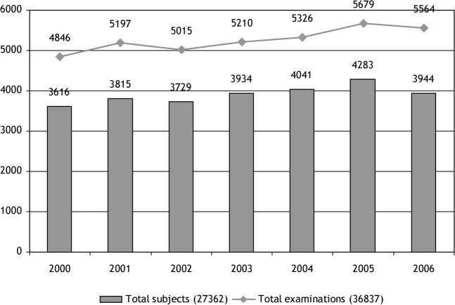
Methods: In 14 major French paediatric radiology departments, children undergoing at least 1 CT scan before age 5, between 2000 and 2006, were included. For each examination, absorbed organ doses were calculated.
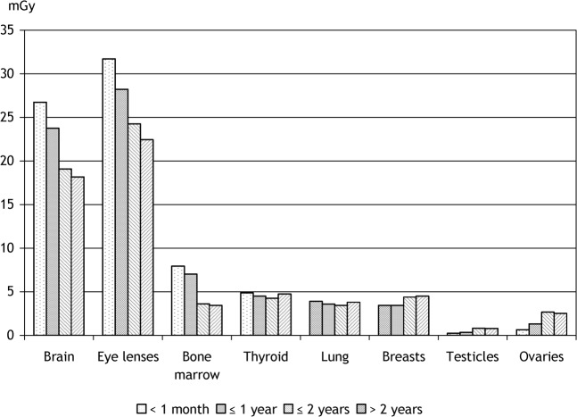
Results: 43% of the 27 362 children in the cohort were aged less than 1 year during their first exposure, with 9% being aged less than 1 month. The mean number of examinations per child was 1.6 (range 1-43). The examinations included: head in 63% of the cases, chest in 21%, abdomen and pelvis in 8% and others in 8%. Brain and eye lenses received the highest cumulative doses from head examinations, with mean organ dose values of 22 mGy (maximum 1107 mGy) and 26 mGy (maximum 1392 mGy), respectively. The mean cumulative effective dose was 3.2 mSv (range 0.1-189 mSv).
Conclusion: CT scan exposure in childhood is responsible for relatively high doses to radiosensitive organs. The rather large dose range according to the protocols used requires their optimisation. The cohort follow-up will study the risk of long-term radiation-induced cancer.
Figures
References
-
- Billon S, Morin A, Caer S, Baysson H, Gambard JP, Backe JC, et al. French population exposure to radon, terrestrial gamma and cosmic rays. Radiat Prot Dosimetry 2005;113:314–20 - PubMed
-
- Boice JD, Jr, Morin MM, Glass AG, Friedman GD, Stovall M, Hoover RN, et al. Diagnostic X-ray procedures and risk of leukemia, lymphoma, and multiple myeloma. JAMA 1991;265:1290–4 - PubMed
-
- Doody MM, Lonstein JE, Stovall M, Hacker DG, Luckyanov N, Land CE. Breast cancer mortality following diagnostic X-rays: findings from the US Scoliosis cohort study. Spine 2000;25:2052–63 - PubMed
-
- Schulze-Rath R, Hammer GP, Blettner M. Are pre- or postnatal diagnostic X-rays a risk factor for childhood cancer? A systematic review. Radiat Environ Biophys 2008;47:301–12 - PubMed
