Case Reports
. 2010 Jun;9(2):186-90.
doi: 10.1007/s12663-010-0057-0. Epub 2010 Sep 28.

Low-grade osteosarcoma of the mandible

Affiliations
Case Reports

Low-grade osteosarcoma of the mandible

Ramen Sinha et al. J Maxillofac Oral Surg. 2010 Jun.

Abstract

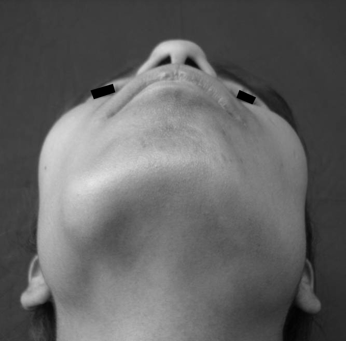
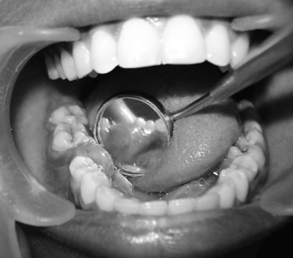
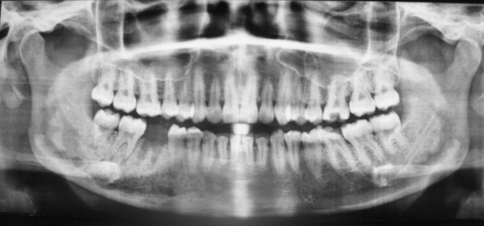
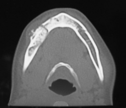
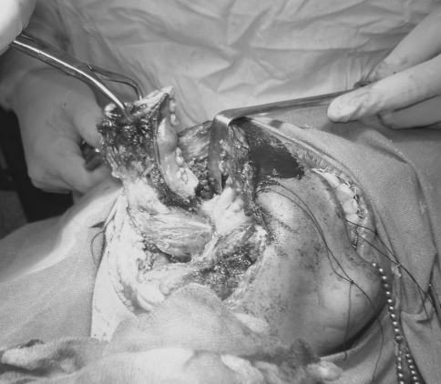
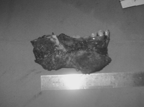
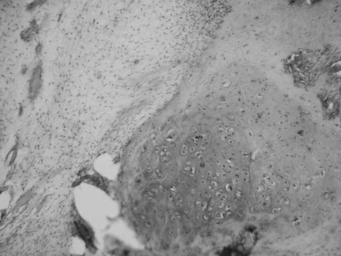
Osteosarcoma (OS), a rare malignant bone tumour arising from primitive bone forming mesenchyme, most often arises in the metaphyses of long bones of the extremities. Bone or osteoid formation within the tumour is characteristic of an osteosarcoma. Craniofacial osteosarcoma (CFOS), most often located in the mandible or maxilla, accounts for only 5-13% of all osteosarcomas. In general, OS of the jaw is a high-grade lesion. Low-grade lesions are rare and represent less than 2% of all osteosarcomas reported in the literature. Because of its rarity and well differentiation, Low-grade OS is usually misdiagnosed as a benign lesion. The clinical and radiographic presentation does not correlate well with the subtle histology picture of a low-grade osteosarcoma which makes the diagnosis difficult.

Keywords: Craniofacial osteosarcoma; Low-grade osteosarcoma.


Figures

Fig. 1
Fig. 1
Extraoral examination showing an ovoid shaped (6 × 4 cm in size) swelling on the right lower border of the mandible
Fig. 2
Fig. 2
Intraoral examination showing a smooth nodular mass in the lower right alveolar ridge extending from 45–48 region
Fig. 3
Fig. 3
Panoramic radiograph showing a diffuse mixed-density lesion, predominating radiopacity interwoven with radiolucent areas, and ill-defined margins involving the right mandible body extending from the 44–48 region
Fig. 4
Fig. 4
Axial CT section showing a mixed density lesion of the right body of the mandible with irregular margins, cortical perforation and soft tissue infiltration
Fig. 5
Fig. 5
Surgical resection of the tumour with safe bony margins of 2 cm
Fig. 6
Fig. 6
Resected specimen
Fig. 7
Fig. 7
Photomicrograph of the tumour showing a variegated appearance with proliferation of small stellate cells and spindle cells in a myxoid matrix. The cells showed moderate nuclear pleomorphism, clumped chromatin and minimal mitotic activity. A few areas of mineralization, chondroblastic differentiation of the osteoid can be seen (H&E ×400)

References

    1. Mardinger O, Givol N, Talmi YP, Taicher S. Osteosarcoma of the jaw. The Chaim Sheba Medical Center experience. Oral Surg Oral Med Oral Pathol Oral Radiol Endod. 2001;91(4):445–451. doi: 10.1067/moe.2001.112330. - DOI - PubMed
    1. Thiele OC, Freier K, Bacon C, Egerer G, Hofele CM. Interdisciplinary combined treatment of craniofacial osteosarcoma with neoadjuvant and adjuvant chemotherapy and excision of the tumour: a retrospective study. Br J Oral Maxillofac Surg. 2008;46(7):533–536. doi: 10.1016/j.bjoms.2008.03.010. - DOI - PubMed
    1. Forteza G, Colmenero B, López-Barea F. Osteogenic sarcoma of the maxilla and mandible. Oral Surg Oral Med Oral Pathol. 1986;62(2):179–184. doi: 10.1016/0030-4220(86)90042-3. - DOI - PubMed
    1. Diniz AF, Filho JA, Alencar Rde C, Garcia RR, Silva MR, Ribeiro-Rotta RF, Silva MA, Batista AC, Mendonça EF. Low-grade central osteosarcoma of the mandible: a case study report. Oral Surg Oral Med Oral Pathol Oral Radiol Endod. 2007;103(2):246–252. doi: 10.1016/j.tripleo.2005.11.006. - DOI - PubMed
    1. Zhao W, Cure J, Castro CY. Low-grade osteosarcoma of the jaw. Ann Diagn Pathol. 2002;6(6):373–377. doi: 10.1053/adpa.2002.36656. - DOI - PubMed

Publication types

LinkOut - more resources