

A comparative evaluation of decalcified freeze dried bone allograft, hydroxyapatite and their combination in osseous defects of the jaws
- PMID: 22190796
- PMCID: PMC3177443
- DOI: 10.1007/s12663-010-0080-1
A comparative evaluation of decalcified freeze dried bone allograft, hydroxyapatite and their combination in osseous defects of the jaws
Abstract
Objectives: To evaluate decalcified freeze dried allograft or hydroxyapatite and a combination of both as bone autograft substitutes in the healing of osseous jaw defects.
Materials and methods: 24 patients participated in the study which involved the filling of osseous defects in the maxilla/mandible with decalcified freeze dried bone allograft (DFDBA) or hydroxyapatite (HA) or a combined graft composed of these two in equal proportions.
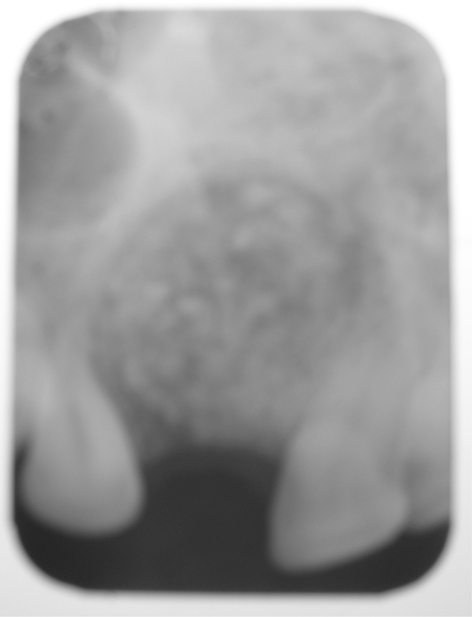
Results: Bone formation occurred as early as 4 weeks in the DFDBA and combination groups and 12 weeks in the HA group which was verified by radiographs, Dentascans (DentaScan® Software Program, General Electric, USA) and bone scintigraphy.
Conclusion: Both these materials can be used as bone graft substitutes in smaller defects although their suitability in large defects is yet to be studied.
Keywords: Bone; Demineralised; Freeze dried; Grafts; Hydroxyapatite.
Figures


References
-
- Putte KA, Urist MR. Osteogenesis in the interior of intramuscular implants of decalcified bone matrix. Clin Orthop Relat Res. 1965;43:257–270. - PubMed
-
- Mupparapu M, Singer SR. Implant imaging for the dentist. J Can Dent Assoc. 2004;70(1):32. - PubMed
-
- Bowen JA, Mellonig JT, Gray JL, Towle HT. Comparison of decalcified freeze-dried bone allograft and porous particulate hydroxyapatite in human periodontal osseous defects. J Periodontol. 1989;60(12):647–654. - PubMed
-
- Mellonig JT. Freeze-dried bone allograft in periodontal reconstructive surgery. Dent Clin North Am. 1991;35(3):505–520. - PubMed
LinkOut - more resources
Full Text Sources