. 2011:2011:903918.
doi: 10.1155/2011/903918. Epub 2011 Dec 10.

Androgen Receptor Regulates Transcription of the ZEB1 Transcription Factor

Affiliations

Androgen Receptor Regulates Transcription of the ZEB1 Transcription Factor

Bynthia M Anose et al. Int J Endocrinol. 2011.

Abstract

The zinc finger E-box binding protein 1 (ZEB1) transcription factor belongs to a two-member family of zinc-finger homeodomain proteins involved in physiological and pathological events mostly relating to cell migration and epithelial to mesenchymal transitions (EMTs). ZEB1 (also known as δEF1, zfhx1a, TCF8, and Zfhep) plays a key role in regulating such diverse processes as T-cell development, skeletal patterning, reproduction, and cancer cell metastasis. However, the factors that regulate its expression and consequently the signaling pathways in which ZEB1 participates are poorly defined. Because it is induced by estrogen and progesterone and is high in prostate cancer, we investigated whether tcf8, which encodes ZEB1, is regulated by androgen. Data herein demonstrate that tcf8 is induced by dihydrotestosterone (DHT) in the human PC-3/AR prostate cancer cell line and that this induction is mediated by two androgen response elements (AREs). These results demonstrate that ZEB1 is an intermediary in androgen signaling pathways.


Figures

Figure 1
Figure 1
Endogenous ZEB1 mRNA expression is induced by DHT. PC-3/AR cells were treated with vehicle (0 nM) or increasing amounts of DHT as indicated. Twenty-four hours later, RNA was harvested and subjected to qPCR using the ZEB1 and RPL32 primers listed in Table 1. ZEB1 mRNA levels were normalized to RPL32 and then plotted relative to the no DHT control. This experiment was repeated 6 times in triplicate. The errors bars represent the standard deviation from the mean average of all experiments. *P < 0.05 compared to no DHT.
Figure 2
Figure 2
Expression of ZEB1 protein is induced by DHT. Protein was isolated from PC-3/AR cells treated with the indicated amounts of DHT for 24 hrs. One hundred mg of protein was used for western blot analysis with a ZEB1 antibody or an α-tubulin antibody as a loading control. The ~150 kDa ZEB protein and the 45 kDa α-tubulin protein are indicated.
Figure 3
Figure 3
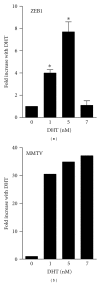
DHT induces tcf8 at the transcriptional level. PC-3/AR cells were transiently transfected with a β-galactosidase reporter plasmid (pBlueZEB974) containing sequences −982 to −9 from tcf8 (left panel) or the MMTV promoter (right panel) and increasing concentrations of DHT as indicated. β-galactosidase activity was determined, and the results are plotted relative to values in the absence of DHT. n = 3, *P < 0.5 compared to no DHT.
Figure 4
Figure 4
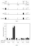
Two functional AREs are present in the tcf8 5′-flanking region. (a) Two well-conserved AREs were identified in tcf8 at −944 and −140 (white boxes) relative to the translation start site. Plasmids were constructed in which each of the putative AREs was mutated individually or both were mutated (black boxes). −36 is the putative transcription start site and is numbered relative to the translation start site. (b). The plasmids depicted in (a) were transfected into PC-3/AR cells with increasing concentrations of DHT as indicated. The cells were harvested after 24 hrs and the β-galactosidase activity in the lysates was determined. *P < 0.05 relative to no DHT. n = 6 with 2-3 replicates per experiment.
Figure 5
Figure 5
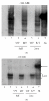
The AREs in tcf8 bind AR. PC-3/AR cells were treated with 5 nM DHT for 24 hrs, and nuclear protein extracts prepared. Oligomers corresponding to the regions of DNA containing (a) the −944 ARE and (b) the −140 ARE were radiolabeled and used as probes. Both GMSAs were run in parallel, with only the ARE probe differing. Ten μg of nuclear protein was used in each lane except for Lane 1, which contained only the radioactive oligomer. Lane 2: plus nuclear extract. Lane 3: plus 50x cold wild-type (WT) self-competitor. Lane 4: plus 50x cold self-competitor with a mutated (MT) ARE. Lane 5: plus 50x cold WT consensus ARE. Lane 6: plus 50x cold MT consensus ARE. Lane 7: plus an AR antibody. The arrow indicates the band representing the AR/DNA complexes supershifted by the antibody.
Figure 6
Figure 6
DHT induces the tcf8 promoter plasmid in a dose-dependent manner in LNCaP cell line derivatives. The LNCaP (a), C4 (b), C4-2 (c), and C4-2B (d) cell lines were transfected with the pBlueZEB974 promoter plasmid and then cultured with increasing DHT as indicated. The cells were harvested after 24 hrs and the β-galactosidase activity in the lysates was determined. (e) C4-2 cells were transfected with the wild type pBlueZEB974 plasmid or with the plasmids containing one or both AREs mutated. Data are plotted relative to the average with no DHT. *P < 0.0001, **P < 0.01, ***P < 0.05.
Figure 7
Figure 7
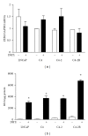
Endogenous tcf8 is not induced in the LNCaP cell lines. The four LNCaP-derived cell lines were each divided into two groups and were incubated with or without 5 nM DHT for 24 hrs. (a) For one group, RNA was isolated and subjected to qPCR for ZEB1 and GAPDH mRNA. ZEB1 mRNA levels were normalized to GAPDH. (b) For the second group, the cells were transfected with pBlueZEB974 at T = O, and β-galactosidase activity was determined at 24 hrs. *P < 0.0001 compared to no DHT.

References

    1. Vandewalle C, Van Roy F, Berx G. The role of the ZEB family of transcription factors in development and disease. Cellular and Molecular Life Sciences. 2009;66(5):773–787. - PMC - PubMed
    1. Chamberlain EM, Sanders MM. Identification of the novel player δEF1 in estrogen transcriptional cascades. Molecular and Cellular Biology. 1999;19(5):3600–3606. - PMC - PubMed
    1. Dillner NB, Sanders MM. Transcriptional activation by the zinc-finger homeodomain protein δEF1 in estrogen signaling cascades. DNA and Cell Biology. 2004;23(1):25–34. - PubMed
    1. Lazarova DL, Bordonaro M, Sartorelli AC. Transcriptional regulation of the vitamin D3 receptor gene by ZEB. Cell Growth and Differentiation. 2001;12(6):319–326. - PubMed
    1. Sekido R, Murai K, Funahashi JI, et al. The δ-crystallin enhancer-binding protein δEF1 is a repressor of E2- box-mediated gene activation. Molecular and Cellular Biology. 1994;14(9):5692–5700. - PMC - PubMed