. 2012 Jan 10;51(1):1-3.
doi: 10.1021/bi2013883. Epub 2011 Dec 28.

Structural analysis of HopPmaL reveals the presence of a second adaptor domain common to the HopAB family of Pseudomonas syringae type III effectors

Affiliations

Structural analysis of HopPmaL reveals the presence of a second adaptor domain common to the HopAB family of Pseudomonas syringae type III effectors

Alex U Singer et al. Biochemistry. .

Abstract

HopPmaL is a member of the HopAB family of type III effectors present in the phytopathogen Pseudomonas syringae. Using both X-ray crystallography and solution nuclear magnetic resonance, we demonstrate that HopPmaL contains two structurally homologous yet functionally distinct domains. The N-terminal domain corresponds to the previously described Pto-binding domain, while the previously uncharacterised C-terminal domain spans residues 308-385. While structurally similar, these domains do not share significant sequence similarity and most importantly demonstrate significant differences in key residues involved in host protein recognition, suggesting that each of them targets a different host protein.


Figures

Figure 1
Figure 1
(A) Cartoon of HopPmaL, HopABPph1448a and AvrPtoB with coloured boxes representing the different structured domains of these proteins. Black lines above the cartoons represent the different fragments used in this study. (B) Ribbon diagram of the HopPmaL[139–207], HopPmaL[281–385] and HopABPph1448a[220–320] structures. Helices are labeled according to the nomenclature of (4).
Figure 2
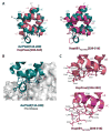
Figure 2
(A) Superposition of AvrPtoB[120–205] (from PDB 3HGK) with HopPmaL[281–385] residues 308–385 (left) and HopAB1Pph1448a[220–320] residues 239–316 (right). N and C represents the N- and C-terminus of AvrPtoB[120–205], while N′ and C′ represents the N- and C-terminus of the superimposed molecule. (B) Closeup of the Pto binding region of AvrPtoB[120–205] as it lies on the surface of the Pto kinase (PDB 3HGK). Selected residues of AvrPtoB[124–200] interacting with Pto kinase are displayed and labeled. (C) Close-up of the putative Pto binding region of HopPmaL[281–385] residues 308–385 and HopAB1Pph1448a[220–320] residues 239–316. Residues from these structures expected to bind Pto based on the AvrPtoB[120–205] structure are shown and labeled.

References

    1. Kim YJ, Lin NC, Martin GB. Cell. 2002;109:589–598. - PubMed
    1. Abramovitch RB, Kim YJ, Chen S, Dickman MB, Martin GB. Embo J. 2003;22:60–69. - PMC - PubMed
    1. Janjusevic R, Abramovitch RB, Martin GB, Stebbins CE. Science. 2006;311:222–226. - PubMed
    1. Dong J, Xiao F, Fan F, Gu L, Cang H, Martin GB, Chai J. Plant Cell. 2009;21:1846–1859. - PMC - PubMed
    1. Xiao F, He P, Abramovitch RB, Dawson JE, Nicholson LK, Sheen J, Martin GB. Plant J. 2007;52:595–614. - PMC - PubMed

Publication types

MeSH terms