. 2012 Jan 17;51(2):686-94.
doi: 10.1021/bi201661g. Epub 2012 Jan 6.

Ionic residues of human serum transferrin affect binding to the transferrin receptor and iron release

Affiliations

Ionic residues of human serum transferrin affect binding to the transferrin receptor and iron release

Ashley N Steere et al. Biochemistry. .

Abstract

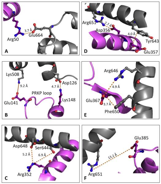
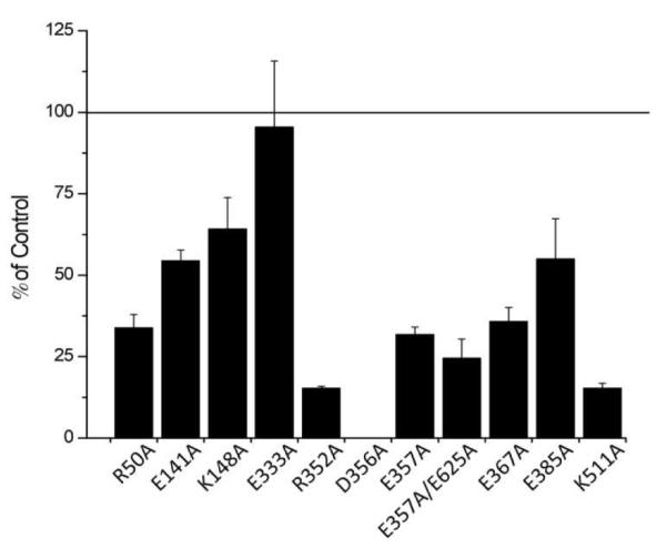
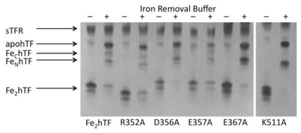
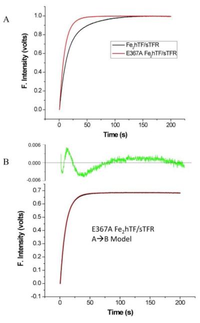
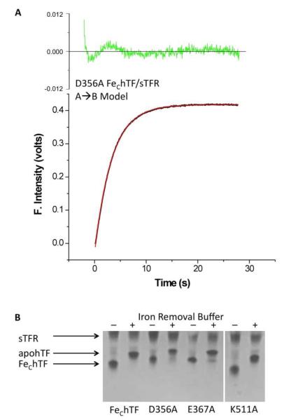
Efficient delivery of iron is critically dependent on the binding of diferric human serum transferrin (hTF) to its specific receptor (TFR) on the surface of actively dividing cells. Internalization of the complex into an endosome precedes iron removal. The return of hTF to the blood to continue the iron delivery cycle relies on the maintenance of the interaction between apohTF and the TFR after exposure to endosomal pH (≤6.0). Identification of the specific residues accounting for the pH-sensitive nanomolar affinity with which hTF binds to TFR throughout the cycle is important to fully understand the iron delivery process. Alanine substitution of 11 charged hTF residues identified by available structures and modeling studies allowed evaluation of the role of each in (1) binding of hTF to the TFR and (2) TFR-mediated iron release. Six hTF mutants (R50A, R352A, D356A, E357A, E367A, and K511A) competed poorly with biotinylated diferric hTF for binding to TFR. In particular, we show that Asp356 in the C-lobe of hTF is essential to the formation of a stable hTF-TFR complex: mutation of Asp356 in the monoferric C-lobe hTF background prevented the formation of the stoichiometric 2:2 (hTF:TFR monomer) complex. Moreover, mutation of three residues (Asp356, Glu367, and Lys511), whether in the diferric or monoferric C-lobe hTF, significantly affected iron release when in complex with the TFR. Thus, mutagenesis of charged hTF residues has allowed identification of a number of residues that are critical to formation of and release of iron from the hTF-TFR complex.


Figures

Figure 1
Figure 1
Location of charged hTF residues in the FeNhTF/sTFR crystal structure (PDB ID: 3S9L)(10). hTF residues (A) Arg50, (B) Glu141 and Lys148, (C) Arg352, (D) Asp356 and Glu357, (E) Glu367, and (F) Glu385 are shown in purple. Residues in the sTFR proposed to interact with the charged hTF residues (Table 1) are shown in gray. Figure generated using PyMOL (18).
Figure 2
Figure 2
Evaluation of the abilities of single point charged-to-alanine Fe2hTF mutants to bind to the sTFR. All mutant hTF samples were prepared at a concentration of 20 μg/mL and competed with biotinylated Fe2hTF for binding to immobilized sTFR. All values are expressed as % of control (Fe2hTF) binding and are averages of at least three different experiments ± standard deviation.
Figure 3
Figure 3
Urea gel analysis of selected charged-to-alanine Fe2hTF mutants in the presence of the sTFR. Samples were electrophoresed before (−) and after (+) incubation with iron removal buffer (100 mM MES, pH 5.6, containing 300 mM KCl and 4 mM EDTA) for 5 min. Note the migration patterns of the charged-to-alanine mutants differ from Fe2hTF due to differences in overall surface charge.
Figure 4
Figure 4
Iron release from the Fe2hTF/sTFR and E367A Fe2hTF/sTFR complexes. (A) Overlay of iron release progress curves from the Fe2hTF/sTFR (black line) and the E367A Fe2hTF/sTFR (red line) complexes. (B) Iron release from the E367A Fe2hTF/sTFR complex (black line) fits best to a simple A→B model (red line). Residuals are shown in green. Attempts to fit the data to alternative models were unsuccessful (Supporting Information Figure 3). All hTF/sTFR samples (375 nM) in 300 mM KCl were rapidly mixed with 200 mM MES, pH 5.6, 300 mM KCl and 8 mM EDTA and excited at 280 nm. Emission was monitored using a 320 nm cut-on filter.
Figure 5
Figure 5
(A) Iron release from the D356A FeChTF/sTFR complex. Iron release from the D356A FeChTF/sTFR complex (black line) fits to a simple A→B model (red line). Residuals are shown in green. The hTF/sTFR sample (375 nM) in 300 mM KCl was rapidly mixed with 200 mM MES, pH 5.6, 300 mM KCl and 8 mM EDTA and excited at 280 nm. Emission was monitored using a 320 nm cut-on filter. (B) Urea gel analysis of selected charged-to-alanine FeChTF mutants in the presence of the sTFR. Samples were electrophoresed before (−) and after (+) incubation with iron removal buffer (100 mM MES, pH 5.6, containing 300 mM KCl and 4 mM EDTA) for 5 min. Note the migration patterns of the charged-to-alanine mutants differ from FeChTF due to differences in overall surface charge.

Similar articles

Cited by

References

    1. Williams J, Moreton K. The distribution of iron between the metal-binding sites of transferrin human serum. Biochem J. 1980;185:483–488. - PMC - PubMed
    1. Huebers H, Josephson B, Huebers E, Csiba E, Finch C. Uptake and release of iron from human transferrin. Proc. Natl. Acad. Sci. U .S. A. 1981;78:2572–2576. - PMC - PubMed
    1. Zak O, Aisen P. Nonrandom distribution of iron in circulating human transferrin. Blood. 1986;68:157–161. - PubMed
    1. Lawrence CM, Ray S, Babyonyshev M, Galluser R, Borhani DW, Harrison SC. Crystal structure of the ectodomain of human transferrin receptor. Science. 1999;286:779–782. - PubMed
    1. Mason AB, Byrne SL, Everse SJ, Roberts SE, Chasteen ND, Smith VC, Macgillivray RT, Kandemir B, Bou-Abdallah F. A loop in the N-lobe of human serum transferrin is critical for binding to the transferrin receptor as revealed by mutagenesis, isothermal titration calorimetry, and epitope mapping. J. Mol. Recognit. 2009;22:521–529. - PMC - PubMed

Publication types