. 2012 Jan;7(1):65-78.
doi: 10.2217/nnm.11.93.

P-glycoprotein silencing with siRNA delivered by DOPE-modified PEI overcomes doxorubicin resistance in breast cancer cells

Affiliations

P-glycoprotein silencing with siRNA delivered by DOPE-modified PEI overcomes doxorubicin resistance in breast cancer cells

Gemma Navarro et al. Nanomedicine (Lond). 2012 Jan.

Abstract

Aims: Multidrug resistance (MDR) mediated by overexpression of drug efflux transporters such as P-glycoprotein (P-gp), is a major problem, limiting successful chemotherapy of breast cancer. The use of siRNA to inhibit P-gp expression in MDR tumors is an attractive strategy to improve the effectiveness of anticancer drugs.

Method: We have synthesized a novel conjugate between a phospholipid (dioleoylphosphatidylethanolamine) and polyethylenimine (PEI) for siRNA delivery, for the purpose of silencing P-gp to overcome doxorubicin resistance in MCF-7 human breast cancer cells.

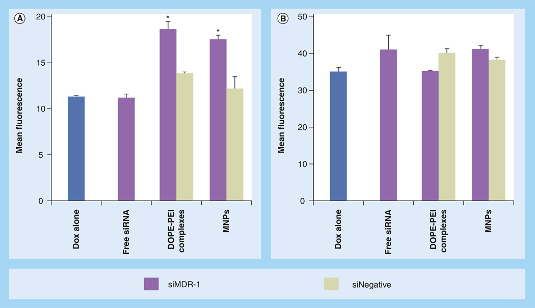
Results: The dioleoylphosphatidylethanolamine-PEI conjugate enhanced the transfection efficacy of low-molecular-weight PEI, which was otherwise totally ineffective. In addition, the polyethylene glycol/lipid coating of the new complexes gave rise to small micelle-like nanoparticles with improved biocompatibility properties. Both coated and noncoated formulations delivered P-gp-specific siRNA to MDR cells.

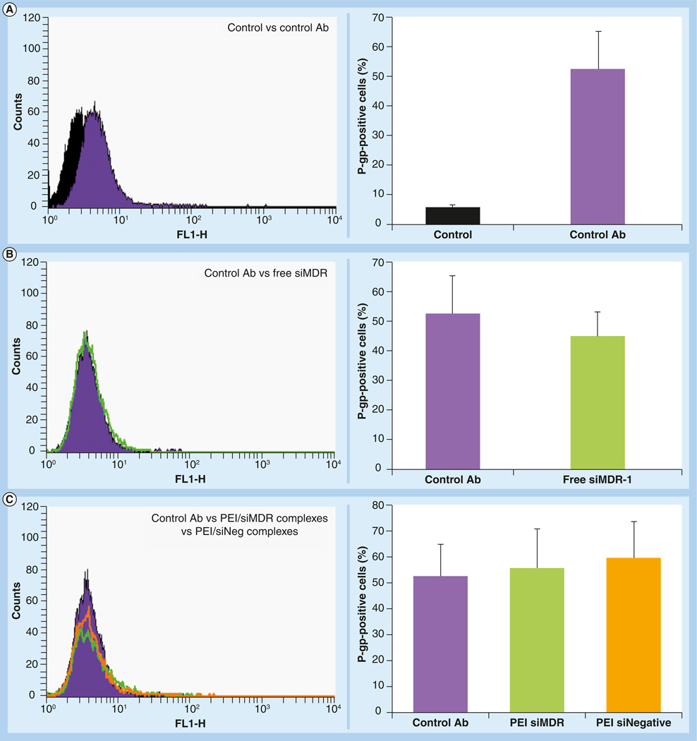
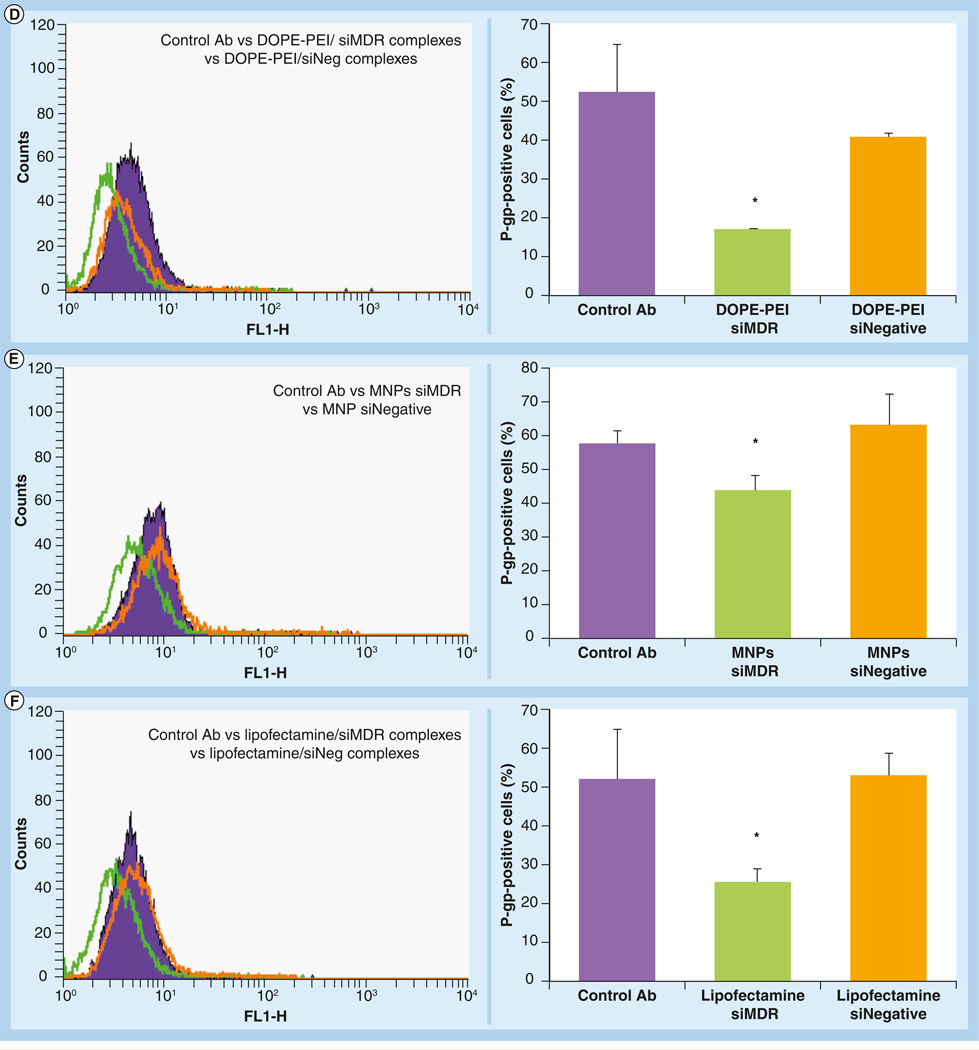
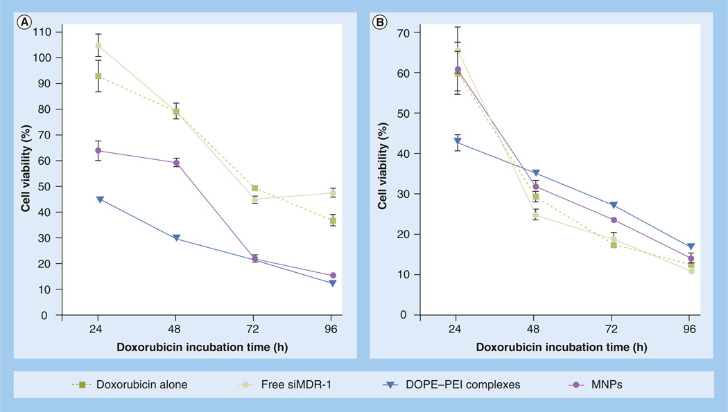
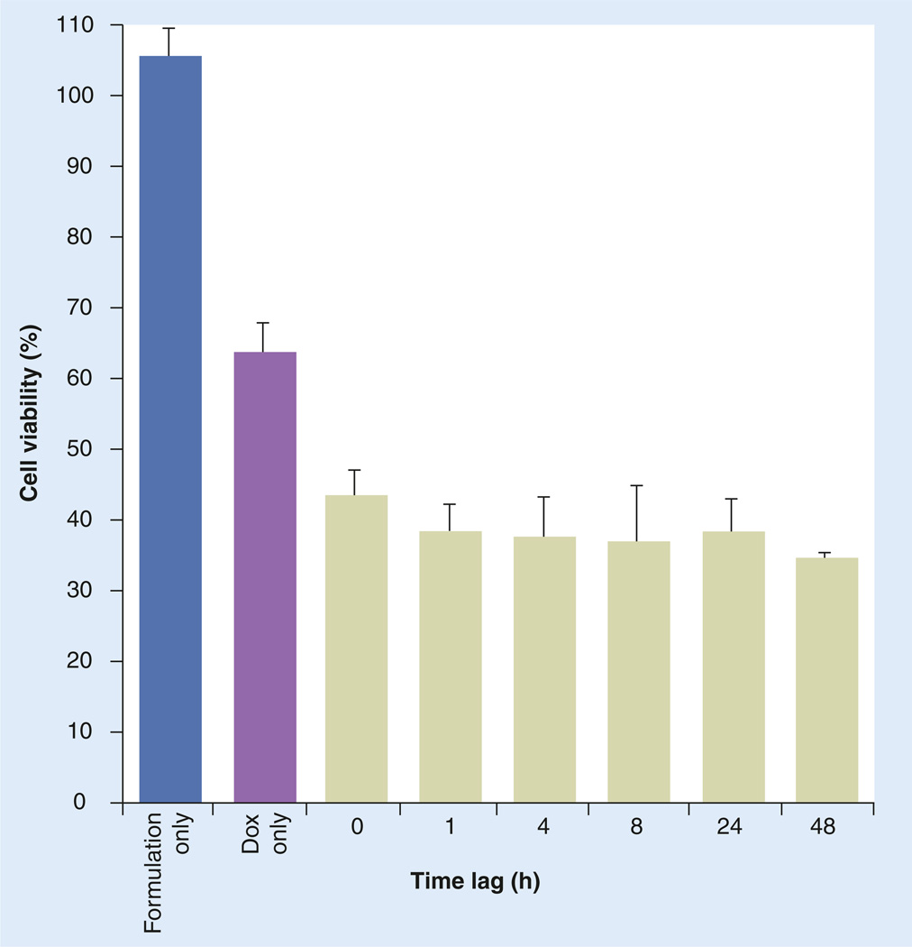
Discussion: The combination of doxorubicin and P-gp silencing formulations led to a twofold increase of doxorubicin uptake and a significant improvement of the therapeutic effect of doxorubicin in resistant cells.


Conflict of interest statement

Financial & competing interests disclosure

The authors have no other relevant affiliations or financial involvement with any organization or entity with a financial interest in or financial conflict with the subject matter or materials discussed in the manuscript apart from those disclosed.

No writing assistance was utilized in the production of this manuscript.

Figures

Figure 1
Figure 1. Green fluorescent protein downregulation by siRNA formulations in c166-green fluorescent protein cells
(A) With PEI 1.8-kDa complexes and (B) DOPE-PEI complexes. c166-GFP cells (stably expressing GFP) were treated with the formulations prepared with GFP-targeted siRNA (n = 3) or nontargeted siRNA (n = 1) at different N/P ratios. The siRNA concentration was 100 nM. After 4 h of incubation, complexes were removed and cells were incubated for 48 h. Cells were trypsinized and analyzed by flow cytometry. The downregulation of GFP was measured by the decrease in the mean fluorescence of the treated cells compared with nontreated and expressed as a percentage of nontreated control cells. DOPE: Dioleoylphosphatidylethanolamine; GFP: Green fluorescent protein; N/P: Polyethylenimine nitrogen/nucleic acid phosphate; PEI: Polyethylenimine.
Figure 2
Figure 2. Effect of polyethylene glycol/lipid layer in micelle-like nanoparticles silencing efficacy
c166-GFP cells (stably expressing GFP) were treated with the formulations prepared with GFP-targeted siRNA or nontargeted siRNA at PEI nitrogen/nucleic acid phosphate ratio of 16. The siRNA concentration was 100 nM. After 4 h of incubation, complexes were removed and cells were incubated for 48 h. Cells were trypsinized and analyzed by flow cytometry. The downregulation of GFP was measured by the decrease in the mean fluorescence of the treated cells compared with nontreated and expressed as a percentage of nontreated control cells. Data are expressed as the mean ± standard deviation (n = 3). DOPE: Dioleoylphosphatidylethanolamine; GFP: Green fluorescent protein; MNP: Micelle-like nanoparticle; PEI: Polyethylenimine.
Figure 3
Figure 3. Decrease in the P-glycoprotein expression in resistant MCF-7 cells mediated by phosphoethanolamine-polyethylenimine complexes and micelle-like nanoparticles
MCF-7 resistant cells were treated with different formulations prepared either with siMDR-1 or siNegative. After 4 h, the treatments were removed and fresh medium was added. The cells were reincubated for 48 h. The cells were detached by mechanical scrapping, resuspended in 0.5% bovine serum albumin, incubated with fluorescein isothiocyanate-labeled antibody against P-gp and analyzed by flow cytometry. Filled histograms correspond to nontreated cells (black, in the absence of antibody and blue in the presence of antibody), green histograms correspond to formulations prepared with siRNA targeting MDR-1 and orange histograms correspond to formulations prepared with scramble siRNA. A shift to the left is indicative of P-gp downregulation. The downregulation of P-gp was quantified by the decrease in mean fluorescence of the treated cells and expressed as a percentage of P-gp positive cells. Data are expressed as the mean ± standard deviation (n = 3). *p < 0.05 versus control cells in the presence of antibody. Ab: Antibody; MDR: Multiple drug resistance; MNP: Micelle-like nanoparticle; P-gp: P-glycoprotein; PEI: Polyethylenimine; siNegative: Scrambled siRNA; siMDR: siRNA targeting MDR-1.
Figure 4
Figure 4. Intracellular doxorubicin levels
In (A) resistant and (B) sensitive MCF-7 cells mediated by DOPE-PEI complexes and MNPs. MCF-7 resistant and sensitive cells were treated with formulations prepared either with siRNA targeting MDR-1 or scramble siRNA. Control cells were treated only with medium. After 4 h, medium was exchanged. Cells were reincubated for 48 h. Cells were then treated for 1 h with doxorubicin (5 µg/ml), then washed, trypsinized and analyzed by flow cytometry. The accumulation of doxorubicin within the cells was measured by the increase in the mean fluorescence. Note ordinate scale differences. Data are expressed as the mean ± standard deviation (n = 3). *p < 0.001 versus Doxalone. DOPE: Dioleoylphosphatidylethanolamine; Dox: Doxorubicin; MNP: Micelle-like nanoparticle; PEI: Polyethylenimine; siNegative: Scrambled siRNA; siMDR: siRNA targeting MDR.
Figure 5
Figure 5. Doxorubicin cytotoxicity
In (A) resistant and (B) sensitive MCF-7 cells after treatment with DOPE-PEI complexes or MNPs. MCF-7 resistant and sensitive cells were treated with formulations prepared with siMDR-1. Control cells were treated with medium. After 4 h, the media was exchanged. Cells were reincubated for 48 h. Cells were treated with doxorubicin (1 µg/ml) for 24, 48, 72 and 96 h and viability was measured. Data are expressed as the mean ± standard deviation (n = 3). p < 0.001 for DOPE-PEI treatments versus drug only treated cells for all time-points in resistant cells. DOPE: Dioleoylphosphatidylethanolamine; MNP: Micelle-like nanoparticle; PEI: Polyethylenimine; siMDR-1: siRNA targeting MDR-1.
Figure 6
Figure 6. Doxorubicin cytotoxicity in resistant MCF-7 cells after treatment with phosphoethanolamine-polyethylenimine complexes at different time lags (h)
MCF-7 resistant and sensitive cells were treated with formulations prepared with siRNA targeting MDR-1 (siMDR-1). Control cells were treated with medium. At different time points post-siMDR-1, cells were treated with doxorubicin (1 µg/ml) for 72 h, and viability was measured. Data are expressed as the mean ± standard deviation (n = 3). p < 0.001 for dioleoylphosphatidylethanolamine-polyethylenimine treatments versus Dox-only treated cells, for all time-points. Dox: Doxorubicin.

References

    1. Gillet JP, Gottesman MM. Mechanisms of multidrug resistance in cancer. Methods Mol. Biol. 2010;596:47–76. - PubMed
    1. Constantinides PP, Wasan KM. Lipid formulation strategies for enhancing intestinal transport and absorption of P-glycoprotein (P-gp) substrate drugs: in vitro/in vivo case studies. J. Pharm. Sci. 2007;96:235–248. - PubMed
    1. Staud F, Ceckova M, Micuda S, Pavek P. Expression and function of p-glycoprotein in normal tissues: effect on pharmacokinetics. Methods Mol. Biol. 2010;596:199–222. ▪ A comprehensive review of the physiological role of P-glycoprotein in normal tissues and its impact on the final fate of drugs.

    1. Marzolini C, Paus E, Buclin T, Kim RB. Polymorphisms in human MDR1 (P-glycoprotein): recent advances and clinical relevance. Clin. Pharmacol. Ther. 2004;75:13–33. - PubMed
    1. Chen H, Hao J, Wang L, Li Y. Coexpression of invasive markers (uPA, CD44) and multiple drug-resistance proteins (MDR1, MRP2) is correlated with epithelial ovarian cancer progression. Br. J. Cancer. 2009;101:432–440. - PMC - PubMed

Publication types

MeSH terms