. 2011 Dec 22:7:101.
doi: 10.1186/1744-8069-7-101.

Rapid S-nitrosylation of actin by NO-generating donors and in inflammatory pain model mice

Affiliations

Rapid S-nitrosylation of actin by NO-generating donors and in inflammatory pain model mice

Jingshan Lu et al. Mol Pain. .

Abstract

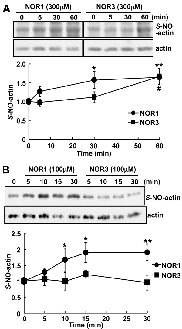
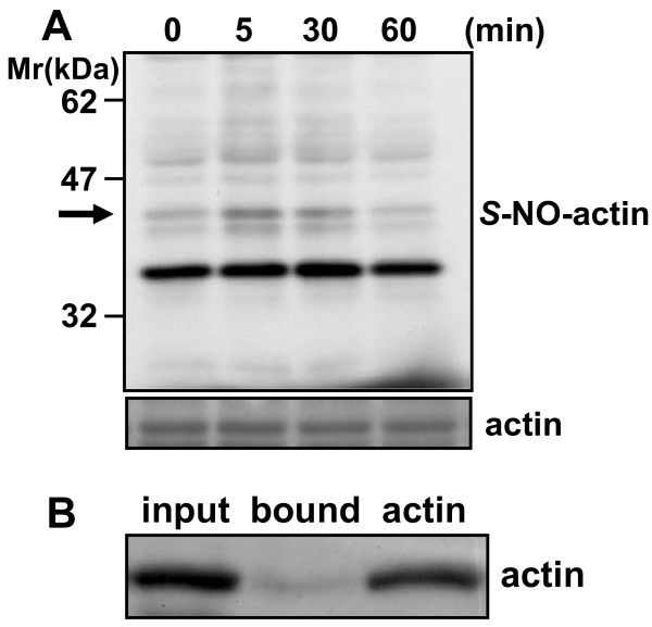
Background: S-Nitrosylation, the reversible post-translational modification of reactive cysteine residues in proteins, has emerged as an important mechanism by which NO acts as a signaling molecule. We recently demonstrated that actin is a major S-nitrosylated protein in the spinal cord and suggested that NO directly attenuates dopamine release from PC12 cells by causing the breakdown of F-actin. However, the occurrence of S-nitrosylation of actin remained unclarified in animal pain model. Kinetic analysis of S-nitrosylation of actin in the present study was made by using NO-generating donors. The biotin-switch assay and purification on streptavidin-agarose were employed for identification of S-nitrosylated actin.

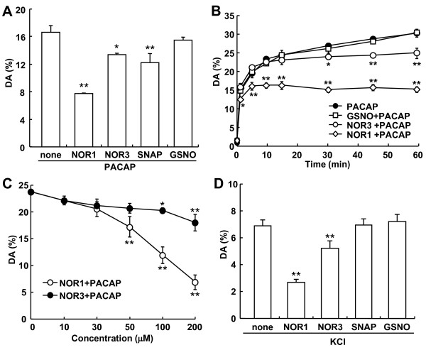
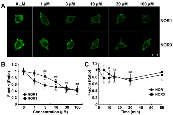
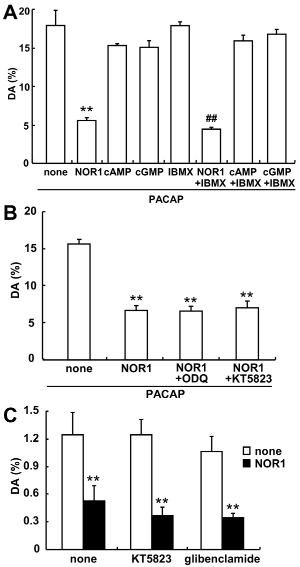
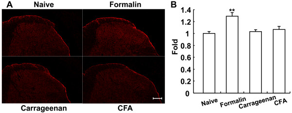
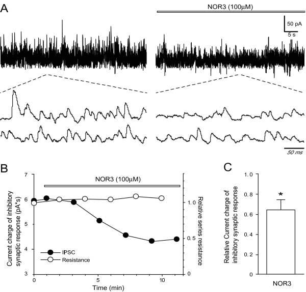
Results: Dopamine release from PC12 cells was markedly attenuated by NOR1 (t1/2 = 1.8 min) and much less by NOR3 (t1/2 = 30 min), but not by S-nitroso-glutathione, an endogenous NO donor. A membrane-permeable cGMP analogue could not substitute for NOR1 as a suppressor nor could inhibitors of soluble guanylate cyclase and cGMP-dependent protein kinase attenuate the suppression. S-Nitrosylated actin was detected by the biotin-switch assay at 5 min after the addition of NOR1. Consistent with the kinetic analysis, actin in the spinal cord was rapidly and maximally S-nitrosylated in an inflammatory pain model at 5 min after the injection of 2% formalin into the hind paws. In vivo patch-clamp recordings of the spinal dorsal horn, NOR3 showed an inhibitory action on inhibitory synaptic transmission in interneurons of the substantia gelatinosa.

Conclusions: The present study demonstrates that rapid S-nitrosylation of actin occurred in vitro in the presence of exogenous NO-generating donors and in vivo in inflammatory pain model mice. Our data suggest that, in addition to the well-known cGMP-dependent protein kinase pathway, S-nitrosylation is involved in pain transmission via disinhibition of inhibitory neurons.


Figures

Figure 1
Figure 1
Effect of NO donors on PACAP-stimulated dopamine release from PC 12 cells. A. Inhibition of PACAP-stimulated dopamine release by NO donors. PC12 cells (4 × 10⁵ cells/well) cultured on 24-well dishes were preincubated for 30 min with NOR1, NOR3, SNAP or GSNO (100 μM concentration of each) and then stimulated with 10 nM PACAP for 5 min in the presence of 10 μM imipramine, an inhibitor of catecholamine reuptake. Dopamine released into the medium and cellular dopamine were measured by HPLC as described in "Methods." *P < 0.05; **P < 0.01 vs. without NO donor. B. Time course of inhibition of PACAP-stimulated dopamine release by NO donors. PC12 cells were stimulated with 10 nM PACAP in the absence and presence of 100 μM NOR1, NOR3 or GSNO, and 10 μM imipramine for the indicated times. *P < 0.05; **P < 0.01 vs. PACAP alone. C. Concentration dependency of NOR1 and NOR3 for the inhibition of PACAP-stimulated dopamine release from PC12 cells. PC12 cells were stimulated for 10 min with 10 nM PACAP in the presence of various concentrations of NOR1 or NOR3 and 10 μM imipramine. Released dopamine (mean ± SD, n = 3) was expressed as a percentage of total dopamine (7.2 ± 0.8 ng/well) in PC12 cells. *P < 0.05; **P < 0.01 vs. without NO donor. D. Inhibition by NO donors of the KCl-stimulated dopamine release in PC12 cells. PC12 cells (4 × 10⁵ cells/well) cultured on 24-well dishes were preincubated for 30 min with NOR1, NOR3, SNAP or GSNO (100 μM concentration of each) in the presence of 10 μM imipramine and then stimulated with 45 mM KCl for 5 min. Dopamine released into the medium (mean ± SD, n = 3) was measured as described above and expressed as a percentage of total dopamine in PC12 cells. **P < 0.01 vs. without NO donor.
Figure 2
Figure 2
Breakdown of F-actin in PC12 cells by NO donors. A. Representative fluorescent images of F-actin in PC12 cells treated with various concentrations of NOR1 or NOR3. PC12 cells (3 × 10⁴ cells/well) were incubated for 5 min with various concentrations of NOR1 or NOR3. Cells were fixed with 4% paraformaldehyde, and labeled with Alexa Fluor 488-phalloidin for F-actin. Bar = 5 μM. B, C. Concentration dependency (B) and time course (C) of F-actin breakdown by NOR1 or NOR3. Fluorescence intensity of more than 40 single cells/dish was quantified by ImageJ as described in "Methods." Data are the mean ± SEM of 3 independent experiments. **P < 0.01 vs. 0 μM NOR1; #P < 0.05; ##P < 0.01 vs. 0 μM NOR3.
Figure 3
Figure 3
No mediation of the cGMP/PKG pathway in inhibition of dopamine release by NOR1. A. Effect of membrane-permeable cGMP and cAMP analogues on PACAP-stimulated dopamine release. PC12 cells (4 × 10⁵ cells/well) cultured on 24-well dishes were stimulated for 10 min with 10 nM PACAP and 100 μM NOR1, 8-Br-cAMP or 8-Br-cGMP in the absence or presence of 100 μM IBMX. **P < 0.01 vs. none; ##P < 0.01 vs. IBMX. B. Effect of inhibitors of the cGMP/PKG pathway on PACAP-stimulated dopamine release by NOR1. PC12 cells were preincubated for 30 min with 100 μM NOR1 and 300 nM ODQ or 1 μM KT5823 in the presence of 10 μM imipramine. Then the cells were stimulated for 5 min by the addition of 10 nM PACAP. **P < 0.01 vs. PACAP alone (none). C. Effect of inhibitors of the cGMP/PKG pathway on basal dopamine release. PC12 cells were incubated for 5 min with 1 μM KT5823 or 1 μM glibenclamide without or with 100 μM NOR1. Dopamine was measured by HPLC, and dopamine released into the medium (mean ± SD, n = 3) was expressed as a percentage of total dopamine in PC12 cells, as described in "Methods." **P < 0.01 vs. none.
Figure 4
Figure 4
S-Nitrosylation of actin by NO donors. A. Time courses of S-nitrosylation of actin in PC12 cells by NO donors. PC12 cells (7 × 10⁵ cells/dish) cultured in 6-cm dishes were exposed to 300 μM NOR1 or NOR3 for the indicated times. The cell lysates in HEN buffer were subjected to the biotin-switch assay as described in "Methods." Samples were resolved by non-reducing SDS-PAGE and immunoblotted with anti-biotin and anti-actin antibodies for S-nitrosylated and total actin, respectively. Intensity of bands was quantified by using ImageJ. The extent of S-nitrosylated actin was normalized to total actin, and the ratio (mean ± SEM, n = 6) of S-nitrosylated actin to total actin at 0 min was taken as "1." *P < 0.05; **P < 0.01 vs. 0 min for NOR1; # P < 0.05 vs. 0 min for NOR3. B. Time courses of S-nitrosylation of actin by NO donors. Purified actin (5 μg) was incubated with 100 μM NO donor NOR1 or NOR3 for the indicated times and subjected to the biotin-switch assay. *P < 0.05; **P < 0.01 vs. 0 min for NOR1.
Figure 5
Figure 5
In vivo S-nitrosylation of actin in the spinal cord of inflammatory pain model. A. Time course of S-nitrosylation of proteins in the spinal cord. The dorsal spinal cords at the L3-L5 levels were dissected at 0, 5, 30 or 60 min after injection of 2% formalin (5 μl) into the hindpaws, and homogenates prepared. The soluble fraction of the homogenates was subjected to the biotin-switch assay. S-Nitrosylated and total actin were detected by anti-biotin and anti-actin antibodies, respectively, as described under "Methods." An arrow indicates the position of S-nitrosylated actin (S-NO-actin). B. Purification of S-nitrosylated actin on streptavidin-agarose. After the soluble fraction prepared from the dorsal spinal cord at 5 min after formalin injection was subjected to the biotin-switch method, S-nitrosylated proteins were purified on streptavidin-agarose. The eluate was resolved on 10% SDS-PAGE, and S-nitrosylated actin was detected with anti-actin antibody. Authentic actin (1 μg) was used as a positive control.
Figure 6
Figure 6
Nitrosocysteine immunoreactivity in the spinal cord of inflammatory pain models. A. Immunoreactivity of nitrosocysteine in the spinal cord. Lumbar transverse sections (20 μm) of spinal cords prepared from before and 5 min, 6 h, and 24 h after injection of formalin, carrageenan, and CFA, respectively, were fixed and stained with anti-nitrosocysteine antibody as described in "Method". Bar = 100 μm. B. Quantification of S-nitrosocysteine immunostaining. Fluorescence intensity of the spinal cord was quantified by ImageJ. The intensity of the spinal cord in inflammatory pain models was normalized to that of naive mice, and the intensity (mean ± SEM, n = 23-24) of naive mice was taken as "1." **P < 0.01.
Figure 7
Figure 7
Representative action of NOR3 on inhibitory synaptic transmission in the SG of the spinal dorsal horn in vivo. A. Spontaneous IPSCs before and 8 min after (left trace) application of NOR3. Two lower consecutive traces of spontaneous IPSCs as shown in an expanded scale in time. B. Time course of the current charge amplitude of spontaneous IPSCs and relative series resistance of the recording under the action of NOR3. Data in A and B were obtained from the same neuron. C. Summary showing inhibitory action of NOR3 on inhibitory synaptic response. *P < 0.05 vs. control.

Similar articles

Cited by

References

    1. Garthwaite J. Concepts of neural nitric oxide-mediated transmission. Eur J Neurosci. 2008;27:2783–2802. doi: 10.1111/j.1460-9568.2008.06285.x. - DOI - PMC - PubMed
    1. Husi H, Ward MA, Choudhary JS, Blackstock WP, Grant SG. Proteomic analysis of NMDA receptor-adhesion protein signaling complexes. Nat Neurosci. 2000;3:661–669. doi: 10.1038/76615. - DOI - PubMed
    1. Meller ST, Gebhart GF. Nitric oxide (NO) and nociceptive processing in the spinal cord. Pain. 1993;52:127–136. doi: 10.1016/0304-3959(93)90124-8. - DOI - PubMed
    1. Ji RR, Kohno T, Moore KA, Woolf CJ. Central sensitization and LTP: do pain and memory share similar mechanisms? Trends Neurosci. 2003;26:696–705. doi: 10.1016/j.tins.2003.09.017. - DOI - PubMed
    1. Hofmann H, Feil R, Kleppisch T, Schlossmann J. Function of cGMP-dependent protein kinases as revealed by gene deletion. Physiol Rev. 2006;86:1–23. doi: 10.1152/physrev.00015.2005. - DOI - PubMed

Publication types