

Myeloid cell tissue factor does not contribute to venous thrombogenesis in an electrolytic injury model
- PMID: 22192154
- PMCID: PMC3336042
- DOI: 10.1016/j.thromres.2011.11.027
Myeloid cell tissue factor does not contribute to venous thrombogenesis in an electrolytic injury model
Abstract
Introduction: Tissue factor (TF) is a potent initiator of the extrinsic coagulation cascade. The role and source of TF in venous thrombotic disease is not clearly defined. Our study objective was to identify the contribution of myeloid cell TF to venous thrombogenesis in mice.
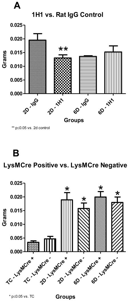
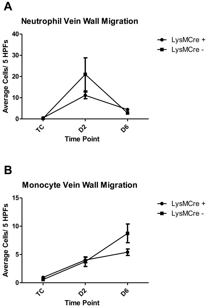
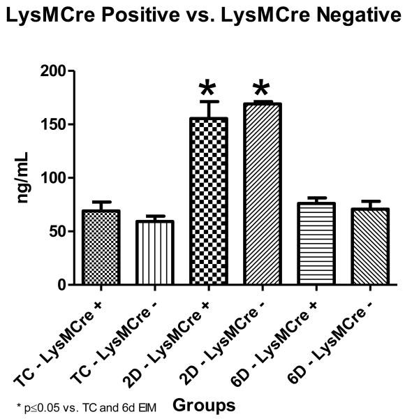
Materials and methods: The mouse electrolytic inferior vena cava model was used to induce thrombosis. The following groups of mice were used (1) TF(flox/flox)LysMCre(+) mice that have reduced TF expression in myeloid cells, (2) TF(flox/flox)LysMCre(-) littermate controls, (3) Wild type mice given a monoclonal anti-mouse TF antibody (1H1) to inhibit TF activity, and (4) Wild type mice given rat IgG. Evaluations at baseline, day 2, and day 6 post thrombosis included thrombus weight, vein wall inflammatory cell migration, vein wall TF mRNA, and plasma D-dimer levels.
Results: Inhibition of TF significantly decreased thrombus weight 2days post venous thrombosis. In contrast, TF(flox/flox)LysMCre(+) had no change in thrombus weight when compared to littermate controls. The absence of myeloid cell TF did not affect infiltration of neutrophils or monocytes into the vein wall. TF mRNA expression in the vein wall decreased at 2days but then returned to baseline levels by 6days post thrombosis. D-dimer levels peaked at 2days post thrombosis in mice with or without myeloid cell TF.
Conclusions: TF is important in the formation of venous thrombi in the macrovasculature. However, TF expression by myeloid cells does not significantly contribute to venous thrombogenesis in this model.
Copyright © 2011 Elsevier Ltd. All rights reserved.
Conflict of interest statement
Figures

Similar articles
-
Inferior vena cava ligation rapidly induces tissue factor expression and venous thrombosis in rats.Arterioscler Thromb Vasc Biol. 2009 Jun;29(6):863-9. doi: 10.1161/ATVBAHA.109.185678. Epub 2009 Mar 5. Arterioscler Thromb Vasc Biol. 2009. PMID: 19265029
-
Male mice have increased thrombotic potential: sex differences in a mouse model of venous thrombosis.Thromb Res. 2011 May;127(5):478-86. doi: 10.1016/j.thromres.2011.01.004. Epub 2011 Feb 5. Thromb Res. 2011. PMID: 21296387 Free PMC article.
-
Circulating and vein wall P-selectin promote venous thrombogenesis during aging in a rodent model.Thromb Res. 2013 Jan;131(1):42-8. doi: 10.1016/j.thromres.2012.10.013. Epub 2012 Nov 20. Thromb Res. 2013. PMID: 23174624
-
Critical review of mouse models of venous thrombosis.Arterioscler Thromb Vasc Biol. 2012 Mar;32(3):556-62. doi: 10.1161/ATVBAHA.111.244608. Arterioscler Thromb Vasc Biol. 2012. PMID: 22345593 Free PMC article. Review.
-
Tissue factor and thrombosis models.Arterioscler Thromb Vasc Biol. 2010 May;30(5):900-8. doi: 10.1161/ATVBAHA.108.177477. Arterioscler Thromb Vasc Biol. 2010. PMID: 20393156 Review.
Cited by
-
Thrombosis: tangled up in NETs.Blood. 2014 May 1;123(18):2768-76. doi: 10.1182/blood-2013-10-463646. Epub 2013 Dec 23. Blood. 2014. PMID: 24366358 Free PMC article. Review.
-
Tumor-derived tissue factor-positive microparticles and venous thrombosis in cancer patients.Blood. 2013 Sep 12;122(11):1873-80. doi: 10.1182/blood-2013-04-460139. Epub 2013 Jun 24. Blood. 2013. PMID: 23798713 Free PMC article. Review.
-
Model-dependent contributions of FXII and FXI to venous thrombosis in mice.J Thromb Haemost. 2020 Nov;18(11):2899-2909. doi: 10.1111/jth.15037. Epub 2020 Aug 28. J Thromb Haemost. 2020. PMID: 33094904 Free PMC article.
References
-
- Heit JA, Cohen AT, Anderson FA Group VTEIA. Estimated annual number of incident and recurrent, non-fatal and fatal venous thromboembolism (VTE) events in the US. Blood. 2005;106:267A–A.
-
- Zhu T, Martinez I, Emmerich J. Venous thromboembolism: risk factors for recurrence. Arterioscler Thromb Vasc Biol. 2009;29:298–310. - PubMed
-
- Himber J, Wohlgensinger C, Roux S, Damico LA, Fallon JT, Kirchhofer D, et al. Inhibition of tissue factor limits the growth of venous thrombus in the rabbit. J Thromb Haemost. 2003;1:889–95. - PubMed
Publication types
MeSH terms
Substances
Grants and funding
LinkOut - more resources
Full Text Sources
Medical
Molecular Biology Databases
Miscellaneous
