Review
. 2012 Jan;28(1):72-86.
doi: 10.1016/j.dental.2011.09.011.

Durability of bonds and clinical success of adhesive restorations

Affiliations
Review

Durability of bonds and clinical success of adhesive restorations

Ricardo M Carvalho et al. Dent Mater. 2012 Jan.

Abstract

Resin-dentin bond strength durability testing has been extensively used to evaluate the effectiveness of adhesive systems and the applicability of new strategies to improve that property. Clinical effectiveness is determined by the survival rates of restorations placed in non-carious cervical lesions (NCCL). While there is evidence that the bond strength data generated in laboratory studies somehow correlates with the clinical outcome of NCCL restorations, it is questionable whether the knowledge of bonding mechanisms obtained from laboratory testing can be used to justify clinical performance of resin-dentin bonds. There are significant morphological and structural differences between the bonding substrate used in in vitro testing versus the substrate encountered in NCCL. These differences qualify NCCL as a hostile substrate for bonding, yielding bond strengths that are usually lower than those obtained in normal dentin. However, clinical survival time of NCCL restorations often surpass the durability of normal dentin tested in the laboratory. Likewise, clinical reports on the long-term survival rates of posterior composite restorations defy the relatively rapid rate of degradation of adhesive interfaces reported in laboratory studies. This article critically analyzes how the effectiveness of adhesive systems is currently measured, to identify gaps in knowledge where new research could be encouraged. The morphological and chemical analysis of bonded interfaces of resin composite restorations in teeth that had been in clinical service for many years, but were extracted for periodontal reasons, could be a useful tool to observe the ultrastructural characteristics of restorations that are regarded as clinically acceptable. This could help determine how much degradation is acceptable for clinical success.


Figures

Fig 1
Fig 1
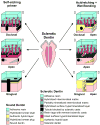
Schematic of potential deterrents to resin-infiltration following total-etching or self-etching in sound and sclerotic dentin of NCCL.
Fig 2
Fig 2
A: This demineralized TEM micrograph showed a hypermineralized layer (HM) within the deepest part of a wedge-shaped lesion that was about 14 μm thick. Bacteria colonies were trapped inside this layer (hollow arrow) by a thin hypermineralized layer (pointer). Another species of bacteria (arrowhead) accumulated along the surface of the hypermineralized layer. Dentinal tubules were not occluded with sclerotic casts and were also filled with bacteria (solid arrow). B: Demineralized TEM micrograph of an “erratic” hybrid layer from the apex of a wedge-shaped lesion that was etched with 40% phosphoric acid and bonded using Clearfil Liner Bond 2V. The thickness of the hybrid layer varied from being absent (arrow) where a hypermineralized layer (HM) was present, to 5 μm (Hd) where the latter was thin and was eroded by bacteria (B). A: adhesive; SD: sclerotic dentin.
Fig 2
Fig 2
A: This demineralized TEM micrograph showed a hypermineralized layer (HM) within the deepest part of a wedge-shaped lesion that was about 14 μm thick. Bacteria colonies were trapped inside this layer (hollow arrow) by a thin hypermineralized layer (pointer). Another species of bacteria (arrowhead) accumulated along the surface of the hypermineralized layer. Dentinal tubules were not occluded with sclerotic casts and were also filled with bacteria (solid arrow). B: Demineralized TEM micrograph of an “erratic” hybrid layer from the apex of a wedge-shaped lesion that was etched with 40% phosphoric acid and bonded using Clearfil Liner Bond 2V. The thickness of the hybrid layer varied from being absent (arrow) where a hypermineralized layer (HM) was present, to 5 μm (Hd) where the latter was thin and was eroded by bacteria (B). A: adhesive; SD: sclerotic dentin.
Fig 3
Fig 3
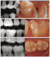
Clinical aspect of resin composite restorations after several years in function, placed by the same operator. They illustrate the satisfactory clinical outcome of earlier adhesives, composite resins and bond techniques in well-motivated, low risk patients. A, B and C correspond to a mesio-occluso-distal resin composite restoration in the first upper left pre-molar after 18 years of clinical service. Materials used were ScotchBond Multi Purpose (3M ESPE) adhesive and Herculite XR (Kerr) resin composite. Generalized wear (B) and marginal staining on the distal, cervical dentin margin (C) can be observed. There were no clinical or radiographic signs of secondary caries; D and E correspond to a mesio-occlusal-buccal resin composite restoration in the first left lower molar after 14 years of clinical service. Materials used were Prime&Bond 2.1 (Dentsply) adhesive and resin composite Charisma (Heraeus-Kulzer). Although significant wear with marginal exposure on the occlusal aspect, no signs of secondary caries were detected both radiographically (D) and clinically (E); F and G are a mesio-occlusal restoration in the first right lower molar and an occlusal restoration in the second right lower molar, both after 7 years of clinical service. Materials applied were Single Bond (3M ESPE) adhesive and resin composite P60 (3M ESPE). No signs of failure due to bond degradation, both radiographically (F) and clinically (G).
Fig 4
Fig 4
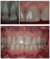
Radiograph, close and wide view of a Class IV transversal fracture restored with UV-cured Nuval-Fil (Dentsply, Caulk) resin composite and using Adaptic ARM (Johnson & Johnson) bonding agent. The photographs and X-Ray were taken on June 2011. The restoration was in service for 35 years. The presence of acid-etched bonded enamel rim ensured retention and marginal integrity of the restoration for many years. (Case treated by Dr. Aymar Pavarini, DDS, PhD, retired Professor of Pediatric Dentistry at Bauru School of Dentistry, University of São Paulo, Bauru, SP, Brazil).
Fig 5
Fig 5
SEM micrographs of laboratory polished/ acid demineralized resin-enamel interface of a retrieved restored tooth after 10 plus years of clinical service. The adhesive joint (AJ) presents a reasonable quality even after such a long period of service in the mouth. Note the loss of silica nanofillers above the adhesive joint detachment in the resin-based composite (RBC) leaving porosities in the adhesive joint; open arrows = filler particle loss.
Fig 6
Fig 6
SEM micrographs of laboratory polished/ acid demineralized adhesive dentin joint of a retrieved restored tooth after 10 plus years of clinical service. A: Adhesive-dentin interface shows good overall morphology with relative small, debonded interface at the bottom of the AJ. It is possible that such defects are artifacts of specimen preparation. Infiltration of the adhesive into dentinal tubules is seen by the present of resin tags both at the adhesive interface and dentinal tubules below it. In this specimen, the tubules can parallel to the dentin surface. B: SEM micrograph of another tooth taken at higher magnification showing large interfacial voids at the middle of the adhesive joint layer but leaving part of the resin adhesive material attached to the underlying sound dentin. This may explain why even in the presence of subclinical interfacial failures there is no dentin sensitivity because the tubules remain sealed with adhesive. The void may be in part due to a thick adhesive joint formed or poor operator performance while applying the adhesive system.

References

    1. Buonocore MG. A simple method of increasing the adhesion of acrylic filling materials to enamel surfaces. J Dent Res. 1955;34:849–53. - PubMed
    1. Brudevold F, Buonocore M, Wileman W. A report on a resin composition capable of bonding to human dentin surfaces. J Dent Res. 1956;35:846–51. - PubMed
    1. Cardoso MV, de Almeida Neves A, Mine A, Coutinho E, Van Landuyt K, De Munck J, Van Meerbeek B. Current aspects on bonding effectiveness and stability in adhesive dentistry. Aust Dent J. 2011;56(Suppl 1):31–44. - PubMed
    1. Pashley DH, Tay FR, Breschi L, Tjaderhane L, Carvalho RM, Carrilho M, Tezvergil-Mutluay A. State of the art etch-and-rinse adhesives. Dent Mater. 2011;27:1–16. - PMC - PubMed
    1. Perdigao J. Dentin bonding-variables related to the clinical situation and the substrate treatment. Dent Mater. 2010;26:e24–37. - PubMed

Publication types