Review
. 2012 Mar;72(3):199-213.
doi: 10.1016/j.neures.2011.12.005. Epub 2011 Dec 17.

Defining and designing polymers and hydrogels for neural tissue engineering

Affiliations
Review

Defining and designing polymers and hydrogels for neural tissue engineering

Emily R Aurand et al. Neurosci Res. 2012 Mar.

Abstract

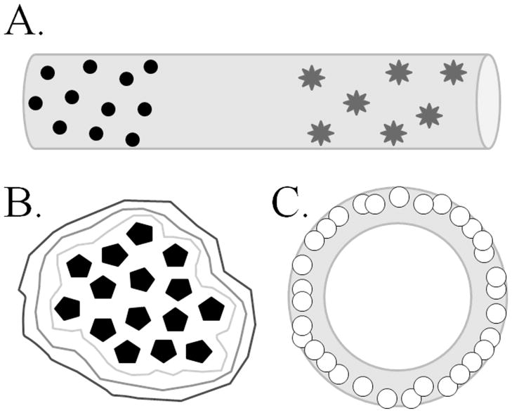
The use of biomaterials, such as hydrogels, as neural cell delivery devices is becoming more common in areas of research such as stroke, traumatic brain injury, and spinal cord injury. When reviewing the available research there is some ambiguity in the type of materials used and results are often at odds. This review aims to provide the neuroscience community who may not be familiar with fundamental concepts of hydrogel construction, with basic information that would pertain to neural tissue applications, and to describe the use of hydrogels as cell and drug delivery devices. We will illustrate some of the many tunable properties of hydrogels and the importance of these properties in obtaining reliable and consistent results. It is our hope that this review promotes creative ideas for ways that hydrogels could be adapted and employed for the treatment of a broad range of neurological disorders.


Figures

Figure 1
Figure 1. Hydrogels can be used to encapsulate cells, microparticles, or therapeutics for multiple delivery purposes
The hydrogel material should be formulated to provide a specific benefit to the encapsulate or host, such as protection from the immune response for encapsulated cells, spatial seclusion of trophic factors, or temporal release of therapeutic compound. (A) Illustrates the use of a hydrogel strand to deliver two different types of microparticles in a spatially- and temporally-defined manner. Lampe, et al. (2011) employed PLGA-based microparticles loaded with BDNF (circles) or GNDF (stars) with differing release kinetics encapsulated within PEG-based hydrogel strands to demonstrate defined release of protein from the hydrogel into the tissue (see text for more details). (B) Wilson, et al. (2008) used the layer-by-layer formulation technique to encapsulate pancreatic islets (pentagons) within layers of different formulations of hydrogel (encompassing lines). The authors fluorescently labeled the different hydrogel formulations to distinguish layers around the islets. The ability to form multiple unique layers of hydrogel around an encapsulate suggests the potential for each layer to contribute a specific, distinct function for both the encapsulate and the host tissue. (C) Using centrifugal casting methods, Mironov, et al. (2005) used HA-based hydrogel seeded with cadiovascular progenitor cells (small circles) to produce hydrogel tubes lined with cells. The overall shape of hydrogel structures, such as tubes or conduits, allow for the production of specialized tissue structures, such as blood vessels or conduits for axon bundles.
Figure 2
Figure 2. Sample parameters for selected hydrogel properties as a function of time (t)
Compressive modulus (solid line) is a measure of the hydrogel strength and increases as the hydrogel polymerizes. During polymerization, cross-links form between monomers, increasing the cross-link density (dotted line). The number of cross-links declines as the bonds are hydrolyzed or cleaved enzymatically during degradation. This also results in a decrease of compressive modulus. The time to degradation (t) is dependent on the chemical composition of the hydrogel and physical properties, such as the incorporation of pores. In most hydrogels, the compressive modulus remains constant during the time between polymerization and degradation. The figure illustrates an example of some sample rates – actual rates depend on a myriad of factors, including chemical and physical properties, incorporations, and external environment.
Figure 3
Figure 3. Survival and fate of peripheral cell types as a function of biomaterial stiffness
This is a general summary of results obtained across a variety of biomaterials, therefore it is important to remember that biomaterial composition can also have an effect on cell survival and proliferation. (A) Chondrocytes appear to grow on a broad range of stiffnesses, however, the differential expression of collagens and ECM components (ie. aggrecan, glycosaminoglycans) could be a function of the stiffness of the biomaterial (Bryant and Anseth, 2002; Nettles et al., 2004; Chung et al., 2006; Lin et al., 2011; Nguyen et al., 2011). (B) Based on available information, fibroblasts may need a less stiff biomaterial for survival and proliferation, compared to studies that have used chondrocytes (Shu et al., 2004; Burdick et al., 2005; Shu et al., 2006). Based on two in vivo studies, encapsulated fibroblasts inserted subcutaneously survive and produce ECM components in biomaterials with both softer and stiffer characteristics (Shu et al., 2004; Shu et al., 2006). (C) Mesenchymal stem cells (MSC) also appear to survive and proliferate better on softer materials, however, changes in stiffness alters the MSC differentiation. In softer materials, MSC begin to express neuronal markers. As the stiffness increases, MSC may begin to express adipogenic or myogenic markers in addition to increasing their expression of factors important to angiogenesis (i.e. VEGF). At the higher range of stiffness, MSC express osteogenic markers (Engler et al., 2006; Chung et al., 2009; Chung and Burdick, 2009; Seib et al., 2009; Huebsch et al., 2010; Quinchia Johnson et al., 2010; Jha et al., 2011).
Figure 4
Figure 4. Survival and fate of neural stem cells as a function of biomaterial stiffness
The fate of neural stem cell (NSC) populations grown on (2-dimensional) or in (3-dimensional) a polymer change as result of varying mechanical properties. Neural cells do not survive well in a biomaterial that is very soft (less stiff) or on a very stiff material. However, those which do survive at the lower stiffness tend towards a neuronal cell fate, whereas astrocytes develop more predominantly at higher stiffness. Neuritic extension is also best observed when the stiffness is lower, but NSC migration is more optimal at slightly higher stiffness (Flanagan et al., 2002; Engler et al., 2006; Mahoney and Anseth, 2006; Saha et al., 2008; Hynes et al., 2009; Lampe et al., 2010a; Lampe et al., 2010b; Seidlits et al., 2010). (A) Post-natal day 6 rat neural stem cells (NSC) developed into >50% neurons, ~25% astrocytes, ~15% oligodendrocytes, and ~10% remained undifferentiated (nestin +) at this approximate level of stiffness (Brännvall et al., 2007). (B and C)Sensory spinal neurons had long neurite extensions but branching was favored on the less stiff material (B) compared to the more rigid material (C). Astrocyte survival was poor on both types of materials (Flanagan et al., 2002). (D–F) Embryonic day 13.5 midbrain-derived NSC, at 24 hours post-encapsulation, had ~54% survival with spheres of mixed populations of neuronal and astrocytic cells (D), ~47% survival (E), and ~31% survival (F), with segregated spheres of neuronal and astrocytic cells. At 21 days, no NSC were surviving in the stiffer hydrogel composition (F) (Seidlits et al., 2010).
Figure 5
Figure 5. Hydrogel pore size changes as a function of precursor molecular weight
Lin, et al. (2011) has demonstrated variation in hydrogel pore size based on the molecular weight of the polymer constituents. Using polyethylene glycol diacrylate (PEGDA) monomer precursors with molecular weights of 3.4kDa (A, box is magnified in a), 6kDa (B, box is magnified in b), 10kDa (C), and 20kDa (D), the authors produced hydrogels with pores (arrows) ranging in size from 9–13μm for hydrogels made from 3.4kDa PEGDA (A), to 38–42μm for 20kDa PEGDA hydrogels (D). The authors observed a similar increase in the nanoscopic property of mesh size depending on molecular weight (45.1Å for 3.4kDa hydrogels and up to 130.9Å for 20kDa hydrogels). These scanning electron microscopy images are of freeze-dried PEGDA hydrogels. It would be unwise to assume that the physical characteristics of the dried hydrogel resemble that of a fully hydrated hydrogel. A fully hydrated hydrogel would be swollen with water and have an amorphous and fibrillary internal meshwork. It is possible that the pore sizes, once hydrated, would be considerable smaller. Scale bar represents 50μm. The above image has been modified from the original by inserting magnification boxes (a, b) to better appreciate the smaller pore sizes. Reproduced with kind permission from Springer Science+Business Media: Pharmaceutical Research, Influence of Physical Properties of Biomaterials on Cellular Behavior, vol. 28, 2011, p.1426, Lin, S., et al.
Figure 6
Figure 6. These PEG-PLA based hydrogels implanted into the rat brain induced a long term neuroimmune response similar to that observed in sham brains penetrated with a needle
Two months after needle penetration (A–C) or hydrogel implant (D–F), a glial response remains. GFAP+ astrocytes (A, D) appear to be more abundant in the tissue surrounding the needle penetration (circled in A) whereas there are fewer astrocytes in the tissue surrounding the hydrogel implant (D). There does appear to be an increase in astrocytes at the hydrogel-brain interface with some GFAP+ processes piercing the hydrogel (D, *). Microglial cells (B, E), identified by positive CD68 reactivity, remain in both the sham brain (B) and in the hydrogel implanted brain (E). While there is not an observable difference in the number of microglia in the surrounding tissues, there is a profusion of microglia that have infiltrated the hydrogel (*). MAP2, labeling identifying neurons (C, F), shows a paucity of MAP2 presence in the area surrounding the needle penetration (circled in C) whereas in the hydrogel brain, MAP2+ cells and neurites can be found closely positioned to the hydrogel (*). Circles in A–C indicate the center of the needle penetration. * in D–F, indicate the center of the hydrogel implant. Arrows (A–C and D–F) indicate corresponding blood vessels found in the adjacent tissue sections. Images were taken between the cerebral peduncle and the subthalamic nucleus. Scale bar indicates 200μm. Quantified data from this study can be found in detail in Bjugstad et al. (2010).

References

    1. Afshari FT, Kappagantula S, Fawcett JW. Extrinsic and intrinsic factors controlling axonal regeneration after spinal cord injury. Expert Rev Mol Med. 2009;11:e37. - PubMed
    1. Ai Y, Markesbery W, Zhang ZM, Grondin R, Elseberry D, Gerhardt GA, Gash DM. Intraputamenal infusion of GDNF in aged rhesus monkeys: Distribution and dopaminergic effects. J Comp Neurol. 2003;461:250–261. - PubMed
    1. Aloisi F. Immune function of microglia. Glia. 2001;36:165–179. - PubMed
    1. Anderson JM, Shive MS. Biodegradation and biocompatibility of PLA and PLGA microspheres. Adv Drug Deliv Rev. 1997;28:5–24. - PubMed
    1. Anseth KS, Bowman CN, Brannon-Peppas L. Mechanical properties of hydrogels and their experimental determination. Biomaterials. 1996;17:1647–1657. - PubMed

Publication types

LinkOut - more resources