. 2012 Mar 15;204(2):389-97.
doi: 10.1016/j.jneumeth.2011.11.031. Epub 2011 Dec 13.

MR-guided stereotactic navigation

Affiliations

MR-guided stereotactic navigation

Shay Ohayon et al. J Neurosci Methods. .

Abstract

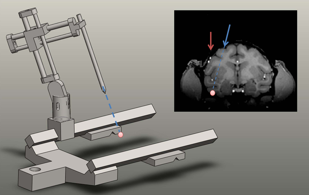
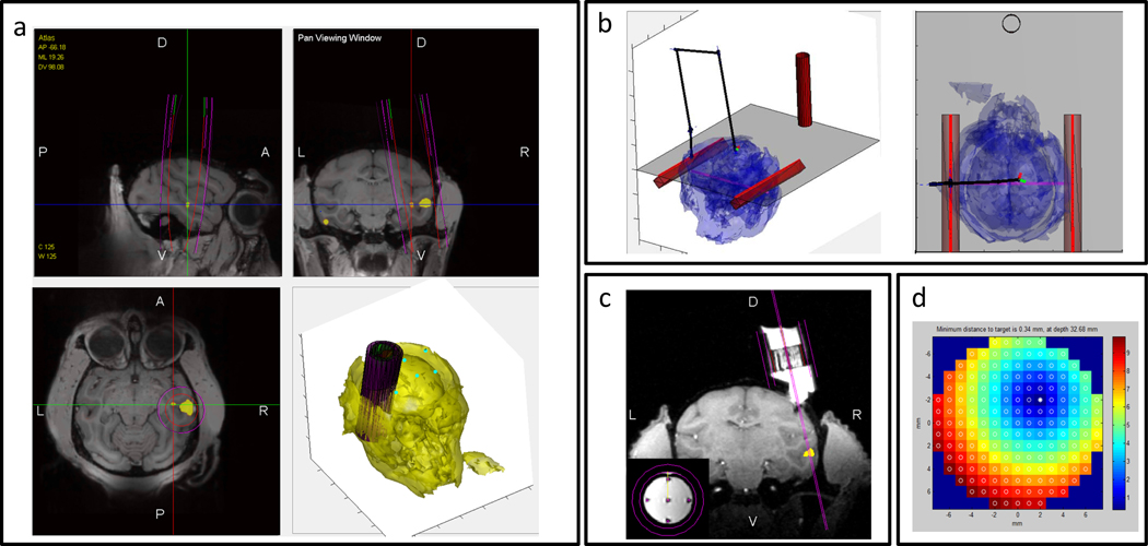
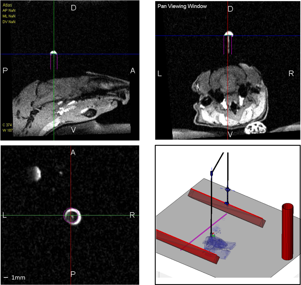
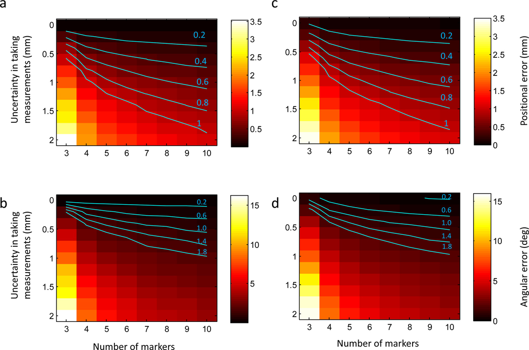
Functional magnetic resonance imaging allows precise localization of brain regions specialized for different perceptual and higher cognitive functions. However, targeting these deep brain structures for electrophysiology still remains a challenging task. Here, we propose a novel framework for MRI-stereotactic registration and chamber placement for precise electrode guidance to recording sites defined in MRI space. The proposed "floating frame" approach can be used without usage of ear bars, greatly reducing pain and discomfort common in standard stereotactic surgeries. Custom pre-surgery planning software was developed to automatically solve the registration problem and report the set of parameters needed to position a stereotactic manipulator to reach a recording site along arbitrary, non-vertical trajectories. Furthermore, the software can automatically identify blood vessels and assist in finding safe trajectories to targets. Our approach was validated by targeting different regions in macaque monkeys and rats. We expect that our method will facilitate recording in new brain areas and provide a valuable tool for electrophysiologists.


Figures

Figure 1.
Figure 1.. The general targeting problem.
A target in an MR scan (pink dot) is selected according to anatomical or functional considerations. The problem is to position the stereotactic manipulator such that the tip aligns with the desired trajectory. Notice that blood vessels above the target site (small white dots) pose a problem for simple vertical penetration (red line), while a non-vertical trajectory can safely reach the target (blue line).
Figure 2.
Figure 2.. Framework overview.
A brain region is selected for targeting and a virtual chamber is placed. Several external markers are rigidly attached to the skull by drilling into an existing implant or securing a small attachment to the head post (not shown). Marker positions in the MR image is identified. During the surgical procedure the position of the markers is read out using the stereotactic manipulator. This can be done even if the stereotactic frame is not physically attached to the animal, but instead to the primate chair. The software solves the targeting problem given the read out values and outputs the set of parameters that are needed to align the manipulator with the planned virtual chamber position.
Figure 3.
Figure 3.. Snapshots from the planning software.
(a) Functional activation map (yellow) is overlaid on top of a structural scan. A virtual chamber is placed (magenta). (b) The stereotactic surgical assistant tool displays the position of the virtual manipulator to reach the desired virtual chamber position. The animal position in the frame is found automatically by registering the markers to their imaged positions. (c) Visualization of the implanted chamber (animal M2) and a virtual electrode to target the desired site. Small inset shows precision of virtual electrodes that align perfectly with five tungsten rods used to fine-calibrate grid rotation (d) Optimal grid analysis automatically finds the best grid tilt angle, orientation and grid hole to use to target the desired site.
Figure 4.
Figure 4.. MR-Guidded cannula placement in a rat.
Snapshots from the planning software demonstrating the planned cannula position (magenta) and the implanted position (white contrast agent).
Figure 5.
Figure 5.. Predicted positional and angular errors of implanting recording chambers.
(a,b) Predicted positional (a) and angular (b) error as a function of the number of external markers and the uncertainty in annotating markers in the MRI scan. (c,d) Predicted errors when noise is present in both MRI marker positions and read out coordinates from the stereotactic manipulator. Iso-error contours are highlighted in cyan.
Figure 6.
Figure 6.. Blood Vessel Avoidance.
(a) Output of the automatic blood detection algorithm in a T1-weighted scan. (b) Coronal view (AP +16) showing two ROIs selected for targeting (denoted as red blobs) and detected blood vessels (highlighted in cyan). Notice that the slice is aligned to stereotactic coordinates and that a major blood vessel in the Superior Temporal Sulcus is present directly above the ROI in the left hemisphere. (c,d) Projected blood patterns on the brain surface. 3D maps were generated by casting rays from the ROI (left, right) and highlighting in red rays that cross through blood vessels. (e) Safe chamber placement selected for targeting the left ROI. (f) View aligned to chamber coordinates. Notice that the electrode track (highlighted in magenta) hits the ROI but does not pass through any blood vessels.

References

    1. Paxinos G, Huang XF & Toga AW The rhesus monkey brain in stereotaxic coordinates. (Academic Press, 2000).
    1. Saleem K Atlas of the rhesus monkey brain in stereotaxic coordinates : a combined mri and histology. 1st edn, (Elsevier, 2006).
    1. Saleem KS & Logothetis N A combined MRI and histology atlas of the rhesus monkey brain in stereotaxic coordinates. (Academic, 2007).
    1. Paxinos G & Watson C The rat brain in stereotaxic coordinates. 6th edn, (Academic Press/Elsevier, 2007).
    1. Sereno MI et al. Borders of multiple visual areas in humans revealed by functional magnetic resonance imaging. Science 268, 889–893 (1995). - PubMed