. 2012 Mar 7;31(5):1241-52.
doi: 10.1038/emboj.2011.471. Epub 2011 Dec 23.

Redox signalling directly regulates TDP-43 via cysteine oxidation and disulphide cross-linking

Affiliations

Redox signalling directly regulates TDP-43 via cysteine oxidation and disulphide cross-linking

Todd J Cohen et al. EMBO J. .

Abstract

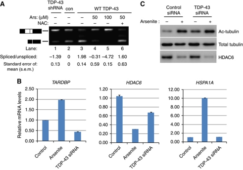
TDP-43 is the major disease protein in ubiquitin-positive inclusions of amyotrophic lateral sclerosis and frontotemporal lobar degeneration (FTLD) characterized by TDP-43 pathology (FTLD-TDP). Accumulation of insoluble TDP-43 aggregates could impair normal TDP-43 functions and initiate disease progression. Thus, it is critical to define the signalling mechanisms regulating TDP-43 since this could open up new avenues for therapeutic interventions. Here, we have identified a redox-mediated signalling mechanism directly regulating TDP-43. Using in vitro and cell-based studies, we demonstrate that oxidative stress promotes TDP-43 cross-linking via cysteine oxidation and disulphide bond formation leading to decreased TDP-43 solubility. Biochemical analysis identified several cysteine residues located within and adjacent to the second RNA-recognition motif that contribute to both intra- and inter-molecular interactions, supporting TDP-43 as a target of redox signalling. Moreover, increased levels of cross-linked TDP-43 species are found in FTLD-TDP brains, indicating that aberrant TDP-43 cross-linking is a prominent pathological feature of this disease. Thus, TDP-43 is dynamically regulated by a redox regulatory switch that links oxidative stress to the modulation of TDP-43 and its downstream targets.


Conflict of interest statement

The authors declare that they have no conflict of interest.

Figures

Figure 1
Figure 1
Oxidative stress regulates TDP-43 subcellular localization and solubility. (A) Cos7 cells were exposed to either 1 mM H2O2 (a–c) or 0.25 mM sodium arsenite (d–f) for 1 h at 37°C in complete media. Cells were subsequently fixed and analysed by immunofluorescence microscopy using rabbit polyclonal TDP-43 and mouse monoclonal TIAR antibodies. Scale bar represents 50 μm. (B) SG localization of either TDP-43 or TIAR in response to H2O2 or arsenite was quantified using 10 fields per image (N=3 independent experiments) and represented as percent TIAR or TDP-43 localization to SGs. P-values were calculated by Student's t-test (***)=2.05 × 10⁻⁵ and (**)=5.1 × 10⁻⁴. Notably, TDP-43 showed preferential localization to H2O2, but not arsenite-induced SGs. (C) Cos7 cells were treated with the indicated oxidative stressors for 1 h and lysates were sequentially extracted with RIPA (R) and urea (U) buffers for analysis by immunoblotting using TDP-43 and GAPDH antibodies. (D) Arsenite-treated cells were washed twice in PBS and allowed a 2-h recovery in arsenite-free complete media followed by immunoblotting analysis as in (C). (E) Cells were pretreated overnight with 20 μg/ml cycloheximide (CHX), where indicated, prior to arsenite exposure and analysed similar to (C) above. Amyloid precursor protein (APP) was used as a positive control for cycloheximide treatment and was detected using an anti-APP-specific antibody (Karen). (F) Cells were treated with arsenite in the presence of 10 mM NAC, where indicated, for subsequent biochemical analysis similar to (C). Figure source data can be found in Supplementary data.
Figure 2
Figure 2
Oxidative stress causes inhibition of TDP-43-mediated RNA regulatory functions. (A) Cells expressing vector control, TDP-43shRNA, or WT TDP-43 plasmids were co-transfected with a CFTR minigene and treated with arsenite and/or NAC where indicated. Spliced and unspliced CFTR transcripts were determined by RT–PCR using specific primers flanking exon 9 followed by gel electrophoresis. RT–PCR products were quantified using the Agilent 2100 Bioanalyzer and relative values of CFTR transcripts are represented as a log ratio of spliced/unspliced products. All samples are normalized to the vector control (lane 2) set to 0. Splicing analysis was performed in triplicate using three independent experiments (N=3), and error bars represent standard error of the mean (s.e.m.). (B) Control siRNA or TDP-43-specific siRNA expressing QBI-293 cells were exposed to 50 μM arsenite overnight, where indicated, to allow full redox-mediated inactivation of TDP-43, and subsequently total RNA was analysed by quantitative RT–PCR using transcript-specific primers for TARDBP, HDAC6, or HSPA1A, which encodes the Hsp70 protein as a control for arsenite exposure. Statistical analysis from N=3 independent replicates was performed using Student's t-test and P-values <2 × 10⁻⁴ for all control versus arsenite and control versus TDP-43 siRNA samples. (C) Control and TDP-43 siRNA samples prepared in parallel similar to (B) above were harvested for protein extraction and western analysis using acetylated tubulin, total tubulin, or HDAC6-specific antibodies. Figure source data can be found in Supplementary data.
Figure 3
Figure 3
TDP-43 cysteine residues undergo oxidation and disulphide bond formation in vitro. (A) Purified recombinant TDP-43 protein was incubated with the indicated concentrations of H2O2 in the presence of [14C]-iodoacetate. Samples were analysed by gel electrophoresis, transferred to nitrocellulose membrane, and developed using a phosphor-imager. (B) Recombinant TDP-43 was treated with 1 mM H2O2 and analysed by non-reducing immunoblotting using a panel of antibodies detecting different TDP-43 epitopes: Ab-1, rabbit polyclonal anti-TDP-43 (Proteintech), Ab-2, polyclonal C-terminal 1038, Ab-3, monoclonal anti-TDP-43 (Proteintech), and Ab-4, polyclonal N-terminal 1065 antibodies. (C) Recombinant TDP-43 treated with H2O2 was subsequently treated with the reducing agent DTT where indicated. All proteins were subsequently cross-linked as a final step using EGS according to the procedures detailed in the Materials and methods section and analysed by immunoblotting using anti-TDP-43 1038. Figure source data can be found in Supplementary data
Figure 4
Figure 4
TDP-43 disulphide bond formation occurs in cultured cells exposed to oxidative stress. (A) QBI-293 cells transfected with a WT TDP-43 expression plasmid were treated with 0.25 mM arsenite for 1 h where indicated. Cell lysates were sequentially extracted and analysed under reducing and non-reducing conditions followed by immunoblotting with anti-TDP-43 1038. Bracketed bands represent HMW TDP-43 species as indicated. (B) Cos7 cell lysates from naive untransfected cells treated with either 0.25 mM arsenite or 10 μM cadmium in the presence of 10 mM NAC, where indicated, were analysed by non-reducing western blotting using anti-TDP-43 1038 or anti-GAPDH antibodies. (C) Urea extracted fractions from arsenite-treated QBI-293 or Cos7 cells were immunoblotted in the presence or absence of DTT and gel mobility of endogenous monomeric TDP-43 was evaluated using anti-TDP-43 1038. (D) Hippocampal neurons isolated from WT C57/BL6 mice were cultured for 10 days prior to treatment with 0.25 mM arsenite for 2 h. Neurons were sequentially extracted similar to (A) and soluble/insoluble lysates were immunoblotted using anti-TDP-43 1038. The asterisk (*) indicates a non-specific cross-reactive band detected with anti-TDP-43 1038 in mouse cells. The bracketed bands highlight HMW TDP-43 species detected under non-reducing conditions. Figure source data can be found in Supplementary data
Figure 5
Figure 5
Oxidative stress induces TDP-43 intra-molecular cysteine interactions as detected by mass spectrometry. (A) Untreated or H2O2 treated (10 mM) recombinant WT TDP-43 were processed in triplicate using non-reducing SDS–PAGE and Coomassie blue staining. HMW multimeric and monomeric TDP-43 species were gel excised for analysis by nanoLC/nanospray/MS/MS. (B) TDP-43 was immunoprecipitated from either soluble or insoluble fractions of arsenite or peroxide-treated cells as described in the Materials and methods section. Immunoprecipitated proteins were analysed by gel electrophoresis and Coomassie staining, and monomeric TDP-43 was gel excised for analysis by nanoLC/nanospray/MS/MS. All data were acquired using a specialized software (Mass Matrix, Case Western University) to detect disulphide bonds. (C) A heat map diagram was generated using Mass Matrix software, which highlights the intra-molecular disulphide-bonded species (C173–C175) identified by mass spectrometry from gel excised TDP-43 protein bands isolated in (A, B). TDP-43 cysteine residues are displayed on x- and y-axes and increasing shades of red indicate significance of disulphide bonding between the indicated residues. Note, only Cys173 and Cys175 undergo highly significant disulphide bond formation. The peptide spectrum and mass peak values corresponding to the disulphide-bonded peptide, WCDCKLPNSK, are shown in Supplementary Figure S3. Figure source data can be found in Supplementary data.
Figure 6
Figure 6
Conserved TDP-43 cysteine residues within and surrounding RRM2 are required for stress-induced disulphide bond formation. (A) A partial TDP-43 sequence alignment from several species is shown. Matched amino-acid residues from the indicated species are shaded in red, conserved cysteine residues are shown in black boxes, and the single non-conserved cysteine residue is boxed in green. (B) Purified recombinant WT, 2CS (C173/175S), or 4CS (C173/175/198/244S) proteins were treated with 1–10 mM H2O2, and subsequently with DTT where indicated, and analysed by non-reducing immunoblotting. The 4CS mutant shows abrogated formation of HMW TDP-43 species in response to H2O2 treatment compared with WT or TDP-2CS proteins. (C) WT, 2CS, or 4CS proteins were similarly immunoblotted for analysis of monomeric TDP-43 gel mobility. Under non-reducing conditions, monomeric WT and 2CS proteins migrated as diffuse TDP-43 species, while the 4CS mutant migrated as a compact monomeric band (lanes 1–3), more similar to the reduced WT protein (lane 4). (D) QBI-293 cells expressing WT TDP-43, the cysteine mutants C173/175S, C198/244S, or the 4CS mutant were treated with arsenite and insoluble fractions were processed in parallel under either reducing or non-reducing conditions followed by immunoblotting with anti-myc (9E10) detecting over-expressed TDP-43 proteins. A darker exposure revealed a significant decrease in HMW TDP-43 bands in the absence of all four cysteine residues. (E) Insoluble lysates from WT and 4CS cell extracts prepared as described in (D) above were analysed by non-reducing immunoblotting using two-fold more 4CS (2.5–10% total insoluble extract) compared with WT (1.2–5% total insoluble extract) to more accurately normalize the monomeric protein input and evaluate formation of HMW TDP-43 species. Figure source data can be found in Supplementary data.
Figure 7
Figure 7
TDP-43 disulphide bond formation occurs in normal and pathological FTLD-TDP brain. (A) Cortical brain sections from control or FTLD-TDP subjects were analysed by immunohistochemistry (IHC) using polyclonal TDP-43 1039, which robustly detects TDP-43 aggregates. Inset represents a × 60 magnification of a characteristic perinuclear TDP-43 inclusion in FTLD-TDP. Scale bar represents 50 μm. (BD) Sequential biochemical extractions of frontal cortex from control or FTLD-TDP subjects was performed and sarkosyl (B) and urea (C, D) fractions were analysed by immunoblotting in parallel under either reducing (lanes 1, 3, 5, and 7) or non-reducing (lanes 2, 4, 6, and 8) conditions using a commercially available polyclonal anti-TDP-43 (B, C) or polyclonal anti-TDP-43 2089 (D), raised against a C-terminal TDP-43 epitope that prominently detects aggregated and ubiquitinated TDP-43 C-terminal fragments. Note, HMW TDP-43 disulphide species bracketed by solid black lines are readily detectable in control subjects (∼90–130 kDa), and more prominently observed in FTLD-TDP subjects (∼90–300 kDa). The reduction of these bands in the presence of DTT was readily observed (B, C), but partially impaired by fibrillar C-terminal aggregated species (D). Figure source data can be found in Supplementary data.
Figure 8
Figure 8
Cysteine-generating ALS mutations produce abnormal disulphide cross-linking in response to oxidative stress. (A) Recombinant purified WT and ALS mutant TDP-43 proteins were left untreated or treated with 10 mM H2O2 for 10 min at 4°C and disulphide banding patterns were analysed by Coomassie staining. The asterisk highlights the ∼90 kDa TDP-43 species specifically produced by the cysteine-generating G348C and S379C mutants. (B) Recombinant TDP-43 proteins were similarly exposed to 1–10 mM H2O2 and analysed by non-reducing immunoblotting using anti-TDP-43 1038. WT, G294A, and R361S proteins showed a typical accumulation of >120 kDa HMW species, while the cysteine-generating mutants showed a unique smeared banding pattern characterized by an ∼90 kDa protein band, as indicated with the asterisk. (C) Cells transfected with WT TDP-43 or a panel of ALS mutations (G348C, S379C, A90V, G294A, or R361S) were treated with 100 μM arsenite for 1 h, sequentially extracted as detailed in the Materials and methods, and insoluble fractions were analysed by non-reducing immunoblotting. Protein bands were visualized using anti-myc 9E10 to detect over-expressed myc-tagged TDP-43 proteins migrating as both monomeric and HMW TDP-43 species. The ∼90 kDa TDP-43 immunoreactive band indicated with the asterisk (*) was detected in G348C and S379C mutants but not control or non-cysteine-generating mutants. Quantification of the 90 and 120 kDa disulphide cross-linked bands is depicted below and was determined by band intensity signals (Multi Guage v2.3) that were estimated as percent disulphide species/total TDP-43 monomer, as assessed on identical image exposures. Figure source data can be found in Supplementary data.

References

    1. Agar J, Durham H (2003) Relevance of oxidative injury in the pathogenesis of motor neuron diseases. Amyotroph Lateral Scler Other Motor Neuron Disord 4: 232–242 - PubMed
    1. Ayala YM, De Conti L, Avendaño-Vázquez SE, Dhir A, Romano M, D'Ambrogio A, Tollervey J, Ule J, Baralle M, Buratti E, Baralle FE (2011) TDP-43 regulates its mRNA levels through a negative feedback loop. EMBO J 30: 277–288 - PMC - PubMed
    1. Ayala YM, Pantano S, D'Ambrogio A, Buratti E, Brindisi A, Marchetti C, Romano M, Baralle FE (2005) Human Drosophila and C. elegans TDP43: nucleic acid binding properties and splicing regulatory function. J Mol Biol 348: 575–588 - PubMed
    1. Ayala YM, Zago P, D'Ambrogio A, Xu YF, Petrucelli L, Buratti E, Baralle FE (2008) Structural determinants of the cellular localization and shuttling of TDP-43. J Cell Sci 121: 3778–3785 - PubMed
    1. Benoit RM, Meisner NC, Kallen J, Graff P, Hemmig R, Cèbe R, Ostermeier C, Widmer H, Auer M (2010) The x-ray crystal structure of the first RNA recognition motif and site-directed mutagenesis suggest a possible HuR redox sensing mechanism. J Mol Biol 397: 1231–1244 - PubMed

Publication types