. 2012 Feb 15;31(4):856-69.
doi: 10.1038/emboj.2011.466. Epub 2011 Dec 23.

A novel GRK2/HDAC6 interaction modulates cell spreading and motility

Affiliations

A novel GRK2/HDAC6 interaction modulates cell spreading and motility

Vanesa Lafarga et al. EMBO J. .

Abstract

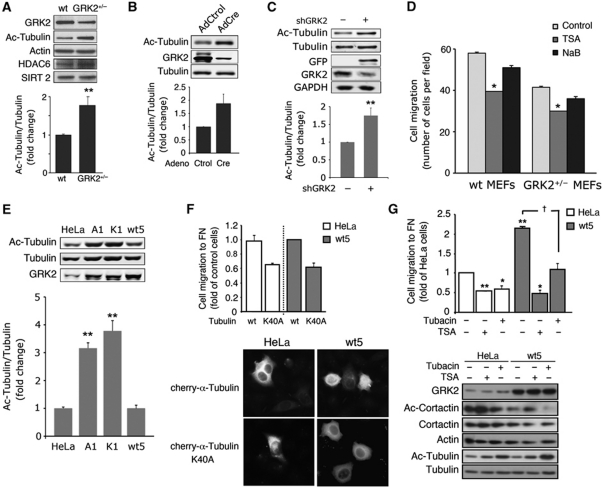
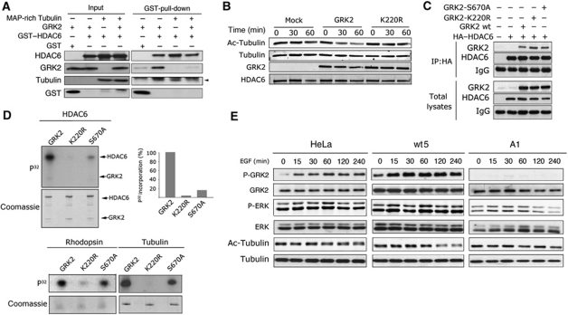
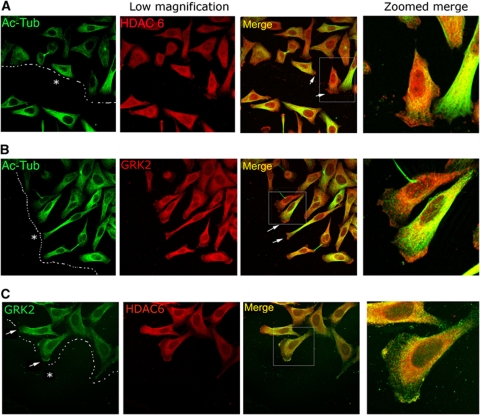
Cell motility and adhesion involves dynamic microtubule (MT) acetylation/deacetylation, a process regulated by enzymes as HDAC6, a major cytoplasmic α-tubulin deacetylase. We identify G protein-coupled receptor kinase 2 (GRK2) as a key novel stimulator of HDAC6. GRK2, which levels inversely correlate with the extent of α-tubulin acetylation in epithelial cells and fibroblasts, directly associates with and phosphorylates HDAC6 to stimulate α-tubulin deacetylase activity. Remarkably, phosphorylation of GRK2 itself at S670 specifically potentiates its ability to regulate HDAC6. GRK2 and HDAC6 colocalize in the lamellipodia of migrating cells, leading to local tubulin deacetylation and enhanced motility. Consistently, cells expressing GRK2-K220R or GRK2-S670A mutants, unable to phosphorylate HDAC6, exhibit highly acetylated cortical MTs and display impaired migration and protrusive activity. Finally, we find that a balanced, GRK2/HDAC6-mediated regulation of tubulin acetylation differentially modulates the early and late stages of cellular spreading. This novel GRK2/HDAC6 functional interaction may have important implications in pathological contexts.


Conflict of interest statement

The authors declare that they have no conflict of interest.

Figures

Figure 1
Figure 1
GRK2 expression levels modulate the extent of tubulin acetylation in MEFs and HeLa cells in a kinase activity-dependent manner. (AC) Downregulation of GRK2 expression enhances tubulin acetylation. MEFs derived from wt or hemizygous GRK2 mice (A), as well as from GRK2-floxed mice infected with control or Cre-recombinase expressing adenovirus (B) or HeLa cells transfected with either a control or a GRK2 silencing construct (C) were lysed and levels of HDAC6, SIRT2, GRK2, tubulin, acetylated tubulin (Ac-tubulin) and actin or GADPH (as loading controls) were determined by western blot analysis. Data of normalized Ac-tubulin levels are mean±s.e.m. from three independent experiments. (D) Motility of MEFs depends on GRK2 expression levels and HDAC6 activity. Cells as in (A) were seeded on Transwell filters precoated with FN (20 μg/ml) in the presence of HDAC inhibitors (TSA, NaB) or vehicle (control). Cell migration was assessed as detailed in Materials and methods. Data are mean±s.e.m. of three independent experiments performed in duplicate. (E) Ac-tubulin markedly accumulates in HeLa cells upon expression of catalytically inactive GRK2 or of the S670A mutant. Parental HeLa cells or cells stably expressing GRK2-wt (wt5), GRK2-S670A (A1) or GRK2-K220R (K1) mutants were analysed for GRK2 levels and the extent of tubulin acetylation determined as above. (F, G) Dynamic deacetylation/acetylation of α-tubulin is involved in the effect of GRK2 on cell migration. Parental and HeLa cells with extra GRK2 were co-transfected with cherry-α-tubulin-wt or cherry-α-tubulin-K40A and the CD-8 antigen (F) or treated (G) with either the general HDAC inhibitor TSA (1 mM) or the HDAC6-specific inhibitor tubacin (15 μM). Transfected tubulin constructs were expressed at similar levels as detected by confocal microscopy (F) and cells positive for co-transfected CD8 were sorted for migration assays by using microbeads precoated with anti-CD8 antibody. Chemotactic motility to FN was assessed as in (D). Data are mean±s.e.m. of three independent experiments performed in duplicate. Total and acetylated levels of α-tubulin and cortactin were measured by western blot (G). Total α-tubulin and actin serve as loading controls. Representative blots are shown in most panels. P<0.05, *P<0.05, **P<0.01, compared with parental, control transfected or infected cells or with vehicle-treated cells, unpaired two-tailed t-test. Figure source data can be found in Supplementary data.
Figure 2
Figure 2
GRK2 associates with and phosphorylates HDAC6 to stimulate tubulin deactetylase activity. (A) HDAC6 co-immunoprecipitates with GRK2. HEK293 cells were transfected with GRK2 alone or together with HA-tagged HDAC6. Protein association was analysed by HA immunoprecipitation followed by immunobloting using anti-HA or anti-GRK2 antibodies. The same antibodies were used to check GRK2 and HDAC6 expression in cell lysates. (B) Association of endogenous HDAC6 and GRK2. Cytoplasmic extracts obtained from HeLa cells were incubated with anti-HDAC6 or IgG antibodies as indicated. Immunoprecipitates (IP) or total cell extracts (input) were analysed by western blot. (C) GRK2 can directly interact with HDAC6. Recombinant GRK2 was incubated with GST alone or with GST-HDAC6 fusion proteins. Proteins bound to Glutathione-sepharose beads were detected with specific anti-GRK2 and anti-GST antibodies. Binding experiments were performed four times with similar results. (D) Identification of the GRK2-binding region in HDAC6. HEK293 cells were transfected with the indicated HA–tagged HDAC6 constructs and co-immunoprecipitation assays performed as above. The expression of HDAC6 constructs or GRK2 in cell lysates was verified as above. Quantification of one of two independent co-immunoprecipitation experiments is shown. (E) HDAC6 is a GRK2 substrate. GRK2 (50 nM) and GST-HDAC6 (50–500 nM) were incubated in the presence of [γ-32P]-ATP as detailed in Materials and methods. The kinetic parameters of the reaction (Vmax and Km) were estimated by double-reciprocal plot analysis. Data are the mean from four independent experiments. (F) A region bearing the second deacetylase catalytic domain of HDAC6 is the main target of GRK2 phosphorylation. HEK293 cells were transfected with the indicated HA-tagged HDAC6 deletion mutants. HA immunoprecipitates were incubated under phosphorylation conditions with recombinant GRK2 (100 nM), followed by SDS–PAGE and autoradiography (upper panel). Overexpression of HDAC6 constructs was monitored by immunoblot. (G) Phosphorylation of HDAC6 by GRK2 enhances tubulin deacetylation. GST-HDAC6 was preincubated with GRK2 or vehicle under phosphorylation conditions, followed by addition of tubulin isolated from TSA-treated HeLa cells. Deacetylase activity was monitored for the indicated times with an anti-acetylated and anti-α-tubulin antibodies. Acetylated band densities were normalized to total tubulin values. A blot representative of two independent experiments is shown. Figure source data can be found in Supplementary data.
Figure 3
Figure 3
Regulation of HDAC6 activity by GRK2 is strictly dependent on its kinase activity and is modulated by GRK2 phosphorylation status. (A) Competition between GRK2 and tubulin for HDAC6 association in vitro. Pull-down assays were performed as in Figure 2C in the presence or absence of purified tubulin. Free and proteins bound to the Glutathione-sepharose beads were immunodetected with specific antibodies. Gels are representative of three independent assays. (B) A catalytically inactive GRK2 mutant is unable to stimulate HDAC6-mediated deacetylation. GST-HDAC6 was preincubated with GRK2, GRK2-K220R or vehicle under phosphorylation conditions, followed by analysis of deacetylation activity as in Figure 2G. GRK2 and HDAC6 levels were monitored to confirm equal loading. (C) GRK2-K220R and GRK2-S670A mutants interact normally with HDAC6. HEK293 cells were co-transfected with HA-tagged HDAC6 in the presence or absence of GRK2-wt or the indicated mutants. GRK2/HADC6 interaction was analysed by co-immunoprecipitation as described in Figure 2A. (D) The GRK2-S670A mutant displays a markedly reduced ability to phosphorylate HDAC6, but not other GRK2 substrates. Phosphorylation of GST-HDAC6 (100 nM), rhodopsin (25 nM) or Tubulin (100 nM) was performed in the presence of [γ-32P]-ATP using recombinant GRK2-wt, GRK2-S670A or GRK2-K220R proteins as described in Materials and methods and Figure 2D. Intensity of 32P-bands was quantified by densitometry and plotted as percentage of wt GRK2-triggered 32P incorporation. Data representative of 2–3 independent experiments are shown. (E) Increased phosphorylation of GRK2 at S670 in response to chemotactic stimuli correlates with active deacetylation of α-tubulin. Parental and HeLa cells stably overexpressing GRK2-wt (wt5) or GRK2-S670A (A1) were challenged with EGF for the indicated times. Levels of acetylated α-tubulin, ERK1/2 activation and the phosphorylation status of GRK2 at S670 were analysed by using specific antibodies as detailed in Materials and methods. Gels are representative of three independent experiments. Figure source data can be found in Supplementary data.
Figure 4
Figure 4
HDAC6 and GRK2 colocalize in the leading edge of migrating cells. HeLa cells stably expressing GRK2-wt were plated in FN (10 μg/ml)-coated dishes and scratched to promote wound healing as indicated in Materials and methods. After 16 h of migration, cells were fixed and potential colocalization of acetylated α-Tubulin with HDAC6 (A) or GRK2 (B) and of HDAC6 with GRK2 (C) was determined by confocal microscopy upon staining with specific antibodies. Arrows indicate the leading edge of migrating cells and dotted lines the margin and direction of the wound. Asterisk denotes wounded area.
Figure 5
Figure 5
GRK2-stimulated HDAC activity is relevant for pseudopodia formation in response to chemotactic cues. (A) Expression of GRK2 mutants unable to phosphorylate HDAC6 inhibits pseudopodia formation. Parental, wt5, A1 or K1 HeLa cells were serum starved for 16 h and subjected to transwell migration assays as detailed in Materials and methods and in the absence or presence of serum in the bottom chamber. Levels of pseudopodia protein recovered on the underside of porous filters were analysed using the Bradford method. Data are mean±s.e.m. of three independent experiments. (B, C) Accumulation of both HDAC6 and GRK2 phosphorylated at S670 at pseudopodia correlates with local deacetylation of tubulin. Cells as in (A) were allowed to migrate in the absence or presence of a serum gradient, and 2 h later purified pseudopodia were collected and the levels of α-Tubulin acetylation (B) or of GRK2, its phosphorylation at S670 and HDAC6 (C) were determined by immunoblot. Data in (B) are mean±s.e.m. from 3 to 4 experiments. Representative blots are shown. *P<0.05, **P<0.01, compared with control, untreated HeLa cells; P<0.05, ††P<0.01 compared with serum-stimulated HeLa cells, unpaired two-tailed t-test. Figure source data can be found in Supplementary data.
Figure 6
Figure 6
Expression of GRK2 mutants defective in HDAC6 regulation results in an altered cell spreading pattern. (A, B) Parental, wt5, A1 or K1 HeLa cells were plated on coverslips coated with FN (10 μg/ml), fixed at the indicated times and analysed by confocal microscopy. The spreading area was quantified by morphometric analysis (A) and cells were triple stained (B) for acetylated α-Tubulin (blue), α-Tubulin (green) and F-actin (Phalloidin, red) as described in Materials and methods. Zoomed images are shown at 20, 40 and 120 min of spreading. Blue, green and white arrows and white arrowheads indicate acetylated MTs, non-acetylated MTs, pioneer MTs, and blebs, respectively.
Figure 7
Figure 7
Enhanced tubulin deacetylation caused by GRK2-mediated HDAC6 phosphorylation modulates cell spreading kinetics and motility. (A) Impairment of GRK2-mediated HDAC6 phosphorylation or pharmacological inhibition of HDAC6 accelerates spreading. The spreading kinetics of parental and HeLa-wt5 pretreated or not with TSA (1 mM) or HeLa-A1 or -K1 cells was analysed using the XCELLigence system as detailed in Materials and methods. (B) Gene-targeted inactivation of GRK2 increases the rate of fibroblast spreading. Primary MEFs derived from GRK2-floxed mice were infected with control or Cre-recombinase expressing adenovirus and their spreading was analysed as above. Total time needed to achieve a maximum cell index during spreading and the extent of tubulin acetylation at this stage was determined for each cell line. Data are mean±s.e.m. of three independent experiments. *P<0.05, **P<0.01, compared with HeLa parental cells or control infected MEFs, unpaired two-tailed t-test. (C) Both the extent and time course of tubulin acetylation during cellular spreading are altered in the presence of GRK2 mutants defective in HDAC6 phosphorylation. Parental and HeLa-wt5, -A1 and -K1 cells were kept in suspension for 2 h and then allowed to adhere and spread into FN-coated plates for the indicated times. Acetylated α-tubulin and total α-tubulin levels were immunodetected with specific antibodies. A representative blot and quantification of tubulin acetylation are shown. (D) Levels of GRK2-pS670 are differentially regulated during spreading and inversely correlate with the spreading rate. Cells were serum starved and collected after kept in suspension (S) or allowed to adhere and spread for 1 h (A) onto FN-coated plates. The extent of GRK2 phosphorylation at S670 and total GRK2 levels were analysed by western blot. A representative blot from two independent experiments is shown. (E) HDAC6-induced migration requires phosphorylation of C-terminal residues on HDCA6 by GRK2. HeLa cells were co-transfected with the CD-8 antigen in the presence of HDAC6-wt or HDAC6-S1060,1062A or S1060,1062,1069A mutants and sorted using microbeads precoated with anti-CD8 antibody. Cell migration was assessed as detailed in Materials and methods. *P<0.05, compared with HDAC6-wt transfected cells (unpaired two-tailed t-test). (F) Expression of a HDAC6 mutant defective in GRK2 phosphorylation accelerates cell spreading kinetics. HeLa cells transfected with GFP-HDAC6-wt or mutant GFP-HDAC6-S1060,1062,1069A were plated on coverslips coated with FN (10 μg/ml), fixed at the indicated times and analysed by confocal microscopy. The spreading area of GFP positive (green labelled) and negative cells was quantified by morphometric analysis and cells were double stained for acetylated α-Tubulin (blue) and F-actin (Phalloidin, red) as in Figure 6. Plotted data are mean±s.e.m. from 10 to 30 cells for each time point and cellular condition. Figure source data can be found in Supplementary data.
Figure 8
Figure 8
Models depicting the intertwinement of GRK2 and HDAC6-mediated tubulin deacetylation in directed cell motility and cellular spreading. (A) Chemotactic movement of cells involves the projection of a dominant cell protrusion in the direction of the chemoattractant source, as a result of localized actin polymerization and the establishment of new adhesions to the substratum. The leading edge is dominated by interrelated structures such as lamella and the organelle-free lamellipodia, which are characterized by different actin and MT networks and distinct extent of focal adhesion (FA) maturation. Association of actin bundles with adhesion sites creates centripetal contractile tension that leads to detachment and retraction of the cell at the rear edge, allowing cell body translocation forward. An increased gradient of MT acetylation is present from the rear to the lamella. This would support cell polarity by facilitating the targeting and dissolution of FA at the rear edge and the delivery of regulatory and structural components to the leading edge. In the lamellipodium, GRK2 would be recruited in a Gβγ-dependent manner to sites of the plasma membrane wherein chemotactic activation is taking place. At such specific locations, chemokine receptor stimulation would promote the phosphorylation of GRK2 at S670 by MAPK, which would in turn switch on the ability of GRK2 to phosphorylate colocalized HDAC6. Phosphorylated HDAC6 would display a higher deacetylase activity towards tubulin at such location, contributing to keep down MT acetylation specifically at the lamellipodium. The presence of highly dynamic, hypoacetylated MTs would stimulate cortical F-actin polymerization by helping to recruit at their plus-ends different small G proteins-GEF activities (that are directly recruited by tubulin or indirectly by the microtubule-interacting +TIP proteins). (B) In the early phase of spreading, hyperacetylation of MTs would increase the rate of spreading as a result of ‘pushing forces’ generated by the sustained growth of stable MT that extend the membrane forward and facilitate the trafficking processes that drive protein cargo to the cell periphery and bring back membrane rafts that were endocytosed during the non-attached, rounded-state of cells before spreading. The extent of bulk MT acetylation would be counterbalanced by the action of HDAC6 in a GRK2-dependent manner. At this stage, local assembly of actin filaments occurs rapidly at the leading membrane edge as integrin contacts with substratum are taking place. MTs entering into this region seem to play a role akin to that of MTs in lamellipodium, displaying lower acetylation levels even in the absence of HDAC6 regulation by GRK2. At later spreading phases, the process turns to rely on FA assembly and actin stress fibres that connect FAs to generate myosin II-dependent traction forces on the substratum. At this phase, hyperacetylation of MTs increases the spreading area by means of stabilization of FA and enhancement of actomyosin contractility, while deacetylated MTs in the lamellipodium contribute to membrane protrusion. In late spreading, the extent of tubulin acetylation of both cortical and non-cortical MTs is determined by the functional interaction of GRK2 with HDAC6.

Similar articles

Cited by

References

    1. Azuma K, Urano T, Horie-Inoue K, Hayashi S, Sakai R, Ouchi Y, Inoue S (2009) Association of estrogen receptor alpha and histone deacetylase 6 causes rapid deacetylation of tubulin in breast cancer cells. Cancer Res 69: 2935–2940 - PubMed
    1. Berzat A, Hall A (2010) Cellular responses to extracellular guidance cues. EMBO J 29: 2734–2745 - PMC - PubMed
    1. Boyault C, Sadoul K, Pabion M, Khochbin S (2007) HDAC6, at the crossroads between cytoskeleton and cell signaling by acetylation and ubiquitination. Oncogene 26: 5468–5476 - PubMed
    1. Cant SH, Pitcher JA (2005) G protein-coupled receptor kinase 2-mediated phosphorylation of ezrin is required for G protein-coupled receptor-dependent reorganization of the actin cytoskeleton. Mol Biol Cell 16: 3088–3099 - PMC - PubMed
    1. Cho SY, Klemke RL (2002) Purification of pseudopodia from polarized cells reveals redistribution and activation of Rac through assembly of a CAS/Crk scaffold. J Cell Biol 156: 725–736 - PMC - PubMed

Publication types