. 2011 Dec 1;124(Pt 23):4027-38.
doi: 10.1242/jcs.087239.

In vivo imaging of basement membrane movement: ECM patterning shapes Hydra polyps

Affiliations

In vivo imaging of basement membrane movement: ECM patterning shapes Hydra polyps

Roland Aufschnaiter et al. J Cell Sci. .

Abstract

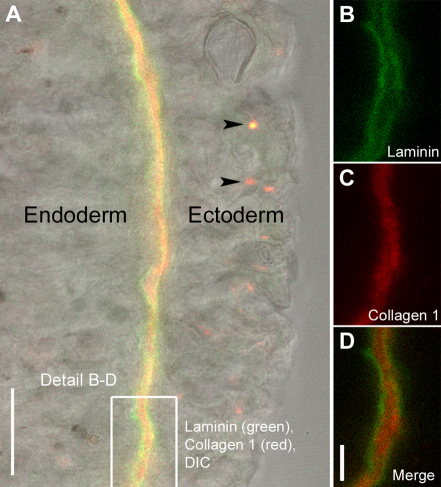
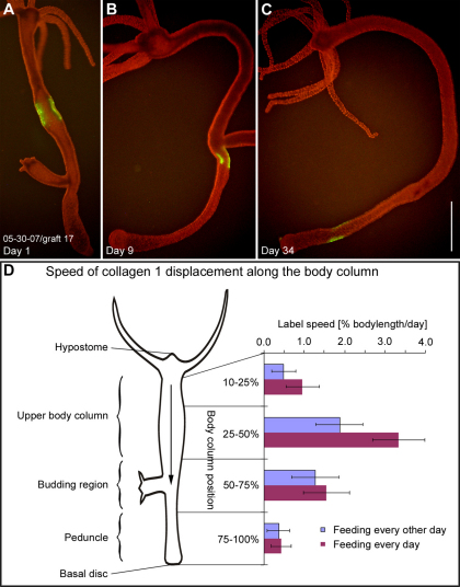
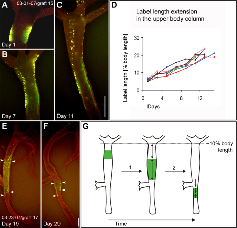
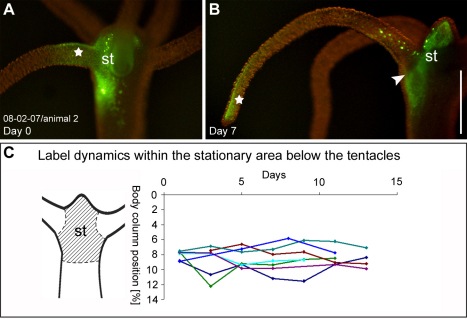
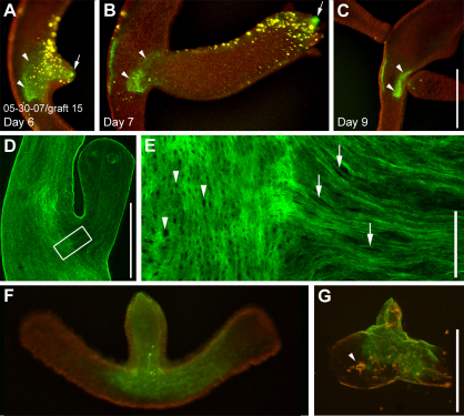
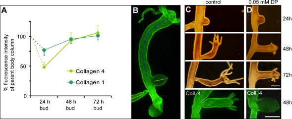
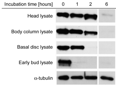
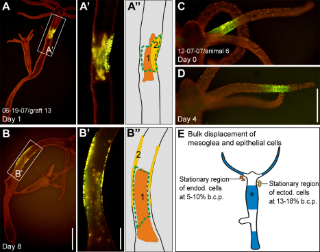
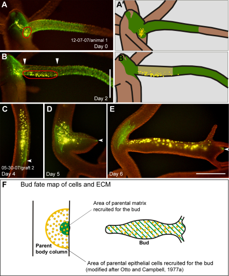
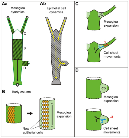
Growth and morphogenesis during embryonic development, asexual reproduction and regeneration require extensive remodeling of the extracellular matrix (ECM). We used the simple metazoan Hydra to examine the fate of ECM during tissue morphogenesis and asexual budding. In growing Hydra, epithelial cells constantly move towards the extremities of the animal and into outgrowing buds. It is not known, whether these tissue movements involve epithelial migration relative to the underlying matrix or whether cells and ECM are displaced as a composite structure. Furthermore, it is unclear, how the ECM is remodeled to adapt to the shape of developing buds and tentacles. To address these questions, we used a new in vivo labeling technique for Hydra collagen-1 and laminin, and tracked the fate of ECM in all body regions of the animal. Our results reveal that Hydra 'tissue movements' are largely displacements of epithelial cells together with associated ECM. By contrast, during the evagination of buds and tentacles, extensive movement of epithelial cells relative to the matrix is observed, together with local ECM remodeling. These findings provide new insights into the nature of growth and morphogenesis in epithelial tissues.


Figures

Fig. 1.
Fig. 1.
Localization of injected antibodies to the three layers of Hydra mesoglea. (A) Optical section through Hydra body wall showing ectoderm, endoderm and the intervening mesoglea in differential interference contrast (DIC) and immunofluorescent labeling. The mesoglea was labeled by injection of monoclonal antibodies mAb52 raised against Hydra laminin (green) and mAb39 raised against Hydra collagen-1 (red). Excess antibody is taken up by ectodermal cells and sequestered in terminal lysosomes (arrowheads). (BD) Labeled mesoglea at higher magnification showing laminin localization in the two subepithelial basal laminae (B,D) and collagen-1 localization in the central interstitial matrix (C,D). Scale bars: 20 μm (A), 5 μm (D).
Fig. 2.
Fig. 2.
Mesoglea displacement along the body column. (AC) A typical graft in the body column labeled for collagen-1 (green) on day 1, day 9 and day 34 after grafting. The labeled area is progressively displaced toward the aboral end of the animal. (D) Mean label displacement speed (x-axis) in different regions of the body column (y-axis). Tip of the hypostome, body column position 0%; basal disc, body column position 100%. Animals fed daily (purple bars) and animals fed every other day (blue bars) were examined. Each bar represents the mean value of individual label velocities (n=10–14) for each body region at each feeding regime. Scale bar: 1 mm.
Fig. 3.
Fig. 3.
Size changes in mesoglea grafts moving down the body column. (AC) A graft labeled for collagen-1 (green) and ectodermal epithelial cells (yellow) in the upper body column at three different time points. The length of the labeled graft increases from 6.6% body length on day 1 to 21% body length on day 11. (D) Length (y-axis) of seven individual grafts labeled for collagen-1 during a time period of 2 weeks (x-axis). The initial length of all grafts is between 5–7% body length and the initial graft position between 10–20% body length on day 1. The length of the labeled grafts was measured every other day. (E,F) A graft labeled for collagen-1 (green) before (E) and after moving through the budding region (F). Note the decrease in size of the labeled area (arrowheads). A decrease in size was typical for all grafts that moved through the budding region. (G) Schematic drawing of labeled graft expansion (1) in the upper body column and labeled graft shrinkage (2) in the budding region during graft displacement along the body column. Scale bar: 500 μm.
Fig. 4.
Fig. 4.
Mesoglea fate in the tentacles and the head region. (A,B) Labeled collagen-1 (green) in the head region and the tentacle base 3 hours after injection and 7 days after injection. The label in the tentacle (star) moves to the tip of the tentacle by day 7, the label in the head region (st) remains stationary. (C) Position (y-axis) of the border between labeled and unlabeled tissue of seven individual head grafts over the course of 11 and 13 days (x-axis). No displacement is observed. Scale bar: 500 μm.
Fig. 5.
Fig. 5.
Mesoglea remodeling during bud formation. (AC) A graft labeled for collagen-1 (green) and epithelial cells (yellow) during bud formation. Tissue labeling preceded bud evagination. At an early stage of evagination, the labeled mesoglea forms a continuous fluorescent patch (A). 24 hours later fluorescence is only visible at the tip (B, arrow) and the base of the bud (B, arrowheads), whereas fluorescence in the intervening region has disappeared. Note the decrease in size of the labeled mesoglea, which remains in the parent body column (A–C, arrowheads). (D) Fixed stage-6 bud stained for collagen-1. (E) Detail from D showing the border region between parent and bud. The collagen-1 matrix in the bud has a clearly stretched appearance: mesoglea pores, which are circular in the parent animal (arrowheads) appear stretched along the oral–aboral axis of the developing bud (arrows). (F) A living bud stage 4–5 labeled for collagen-1 (green). (G) Isolated mesoglea of the same bud as in F. Some endodermal cell debris (arrowhead) remains trapped in the ECM. Scale bars: 500 μm (C,G), 50 μm (E).
Fig. 6.
Fig. 6.
The role of collagen synthesis during bud formation. (A) Relative protein concentration of collagen-1 and collagen-4 in different bud stages. The graph shows average fluorescence intensities of collagen-1 and collagen-4 in 24 hour, 48 hour and 72 hour buds (n=10 for each time point). (B) For the values in the graph, the fluorescence intensity of immunocytochemically labeled specimens was measured along a line in the center of the bud (yellow lines) and compared with the fluorescence intensity of the upper body column of the parent animal (blue line). (C) Control bud development at 24, 48 and 72 hours and a 48 hour control bud stained for collagen-4. (D) A typical example of bud development under continuous treatment with 0.05 mM DP and a 48-hour-treated bud stained for collagen-4. Scale bar: 500 μm.
Fig. 7.
Fig. 7.
Mesoglea turnover in different body regions estimated as laminin stability in tissue lysates. Isolated mesoglea samples containing laminin were incubated in tissue lysates from different body regions of the animal as indicated. For head lysate, only the head without tentacle tissue was used (compare Fig. 4C, st). Bud lysate was taken from bud stages 3–5. Samples taken at different time points were analyzed by western blotting using antibodies against laminin and α-tubulin. The α-tubulin is not significantly degraded in any of the lysate samples; the α-tubulin blot shown corresponds to the basal disc lysate.
Fig. 8.
Fig. 8.
Epithelial cell and mesoglea dynamics in the body column and tentacles. Representative examples of labeled tissue displacement in individual animals. (A,B) A graft labeled for collagen-1 (green), ectodermal epithelial cells (bright yellow) and endodermal epithelial cells (orange) in the upper body column on day 1 and day 8. (A′,B′) Magnified views. (A″,B″) Schematic views. The perspective of A is about 180° rotated from B, as indicated by the position of cell cluster 2 in A″ and B″. (C,D) Labeled patch of collagen-1 (green) and ectodermal epithelial cells (yellow) in a tentacle on day 0 and day 4. Cells and ECM retain a 100% overlap. (E) Results of mesoglea and cell sheet movement illustrated schematically based on observations in multiple animals (n=10 in head and tentacle base, n=10 in tentacle, n=30 in the upper body column, n=50 in budding area and bud, n=15 in the lower peduncle region). Regions where epithelial cells and mesoglea move together (blue) are shown. In the upper body column, the ectodermal layer moves slightly slower than the endodermal layer and the mesoglea (asterisk). The stationary region for ectodermal epithelial cells is located at a body column position of about 13–18% (yellow double-headed arrow) and for endodermal epithelial cells at a body column position of about 5–10% (orange double-headed arrow). Scale bars: 1 mm (B), 500 μm (B,D).
Fig. 9.
Fig. 9.
Epithelial cell and mesoglea dynamics at the tentacle base and during budding. (A) Head region of an animal with labeled mesoglea (green) extending from the head region into the tentacle and a group of labeled ectodermal epithelial cells (yellow, circled in red) between two tentacles at day 0. (A′) Schematic. (B) On day 2, the group of epithelial cells (circled in red) have moved from the stationary head region into the tentacle. Note the gap with strongly reduced green fluorescence at the proximal end of the tentacle (arrowheads) indicating mesoglea expansion. (B′) Schematic. (CE) A graft labeled for collagen-1 (green) and epithelial cells (yellow) in the body column over the course of three consecutive days before (C) and during (D,E) budding. Cells move relative to their underlying substrate into the newly developing animal (D,E). Note that only a small part of the labeled, parental mesoglea is recruited into the bud (arrowheads). (F) Fate map of epithelial cells and ECM of the parent body column recruited into a bud. The schematic shows the area of epithelial cells recruited to the bud (yellow) compared with the area of ECM recruited to the bud (green). The fate map was constructed based on an idealized body column diameter (Otto and Campbell, 1977a). Scale bars: 500 μm.
Fig. 10.
Fig. 10.
Fate map of Hydra mesoglea and a model of cell–matrix interactions during growth and morphogenesis. (A) Mesoglea (a) and epithelial cell (b) dynamics. Black arrows in a indicate the direction of mesoglea movements; shaded areas indicate mesoglea stretching and growth; white areas indicate stationary mesoglea in the head and bud tip; stars mark sites of mesoglea degradation. Yellow arrows in b indicate the direction of epithelial cell movements; shaded area marks the area of epithelial cell proliferation (Campbell, 1967a; Holstein et al., 1991). (B) Mesoglea growth in the body column is associated with cell proliferation. (C) Mesoglea stretching (top) and cell movements (bottom) during steady state tentacle evagination. Light green represents the area of mesoglea expansion (double-headed arrow); dark green represents the region of stable mesoglea; arrow indicates the direction of cell movement. The relationship between cells and substrate changes during these morphogenetic movements: cells move over stable mesoglea (1); cells move over expanding mesoglea (2); cells moved together with mesoglea (3). (D) Expansion of the mesoglea (top) and cell movements (bottom) during bud evagination. Labeling analogous to that in C.

References

    1. Benazeraf B., Francois P., Baker R. E., Denans N., Little C. D., Pourquie O. (2010). A random cell motility gradient downstream of FGF controls elongation of an amniote embryo. Nature 466, 248-252 - PMC - PubMed
    1. Bertet C., Sulak L., Lecuit T. (2004). Myosin-dependent junction remodelling controls planar cell intercalation and axis elongation. Nature 429, 667-671 - PubMed
    1. Bosch T. C., David C. N. (1984). Growth regulation in Hydra: relationship between epithelial cell cycle length and growth rate. Dev. Biol. 104, 161-171 - PubMed
    1. Burnett A. L., Hausman R. E. (1969). Mesoglea of Hydra. 2. Possible Role in Morphogenesis. J. Exp. Zool. 171, 15-24 - PubMed
    1. Campbell R. D. (1967a). Tissue dynamics of steady state growth in Hydra littoralis. I. Patterns of cell division. Dev. Biol. 15, 487-502 - PubMed

Publication types

MeSH terms