Review
. 2011:76:1-16.
doi: 10.1101/sqb.2011.76.010462. Epub 2011 Dec 22.

Bioenergetic origins of complexity and disease

Affiliations
Review

Bioenergetic origins of complexity and disease

D C Wallace. Cold Spring Harb Symp Quant Biol. 2011.

Abstract

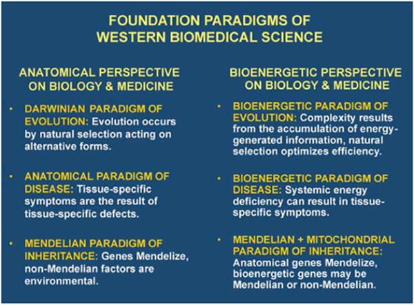
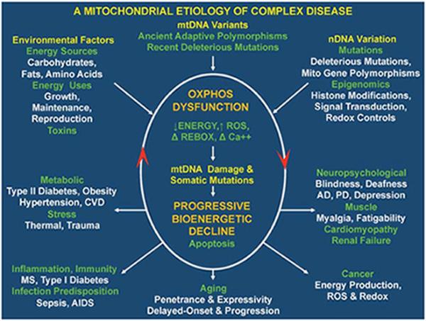
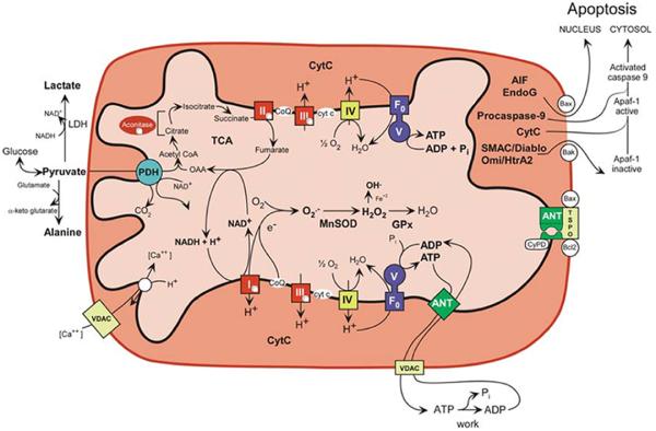
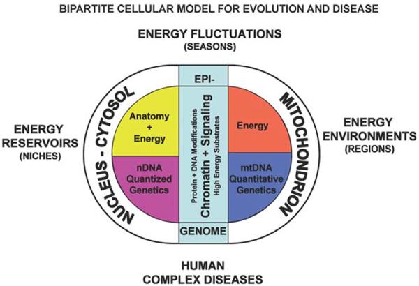
The organizing power of energy flow is hypothesized to be the origin of biological complexity and its decline the basis of "complex" diseases and aging. Energy flow through organic systems creates nucleic acids, which store information, and the annual accumulation of information generates today's complexity. Energy flow through our bodies is mediated by the mitochondria, symbiotic bacteria whose genomes encompass the mitochondrial DNA (mtDNA) and more than 1000 nuclear genes. Inherited and/or epigenomic variation of the mitochondrial genome determines our initial energetic capacity, but the age-related accumulation of somatic cell mtDNA mutations further erodes energy flow, leading to disease. This bioenergetic perspective on disease provides a unifying pathophysiological and genetic mechanism for neuropsychiatric diseases such as Alzheimer and Parkinson Disease, metabolic diseases such as diabetes and obesity, autoimmune diseases, aging, and cancer.


Figures

Figure 1
Figure 1
The anatomical and bioenergetic complementarities in evolution, medicine, and genetics. Comparison of the three pairs of complementary paradigms derived from the anatomical perspective and the bioenergetic perspective.
Figure 2
Figure 2
Integrated mitochondrial paradigm to explain the genetic and phenotypic complexities of the “complex” human diseases. An integrated model for the genetics and pathophysiology of complex diseases, aging, and cancer. The top of the figure indicates the three types of variation that impact individual mitochondrial oxidative phosphorylation (OXPHOS) robustness and hence risk for developing disease symptoms. These include nuclear DNA (nDNA) variation encompassing DNA sequence changes and epigenomic modification of gene regulation and signal transduction pathways, mitochondrial DNA (mtDNA) variation including recent deleterious mutations and ancient adaptive polymorphisms, and environmental influences encompassing the availability and demand for calories and inhibition of mitochondrial function by environmental insults. The central oval encompasses the pathophysiological basis of disease processes and the basis of disease progression. The primary defect is reduction in the energy transformation capacity of OXPHOS. This can result in reduced energy output, increased reactive oxygen species (ROS) production, altered redox status, and altered calcium homeostasis. The decline in OXPHOS efficiency can in turn perturb mitochondrial biogenesis, increase ROS production, impair mitophagy, and so on, resulting in the progressive increase in mtDNA damage and somatic mutations and further decline in mitochondrial function. Once mitochondrial function falls below the bioenergetic threshold of a tissue, symptoms ensue. Continued energetic failure can initiate cell destruction by apoptosis or necrosis. The lower five derived disease categories summarize the phenotypic outcomes of perturbed mitochondrial energy transformation. The bottom arrow shows the effect of the stochastic accumulation of somatic mtDNA mutations, resulting in delay-onset and progressive course of diseases and aging. The right arrow indicates clinical problems that can result from reduced energy production in the most energetic tissues, the brain, heart, muscle, and kidney. The number and severity of symptoms in these organs reflect the degree and specific nature of the mitochondrial defect. The arrow to the left indicates the metabolic effects of mitochondrial dysfunction that result in the perturbation of the body's energy balance. This results in the symptoms of the metabolic syndrome. The arrow toward the lower right corner indicates mitochondrial alterations that are critical for cancer initiation, promotion, and metastasis. The arrow to the lower left corner outlines the hypothesized inflammatory and autoimmune responses that may result from the chronic introduced into the bloodstream of the mitochondria's bacteria-like DNA and N-formyl methionine proteins.
Figure 3
Figure 3
Mitochondrial energy production and its reaction to the pathophysiology of disease. Five features of mitochondrial metabolism are central to the pathophysiology of the common age-related diseases: (1) energy production by OXPHOS; (2) regulation of cellular oxidation–reduction (redox) state; (3) ROS generation as a by-product of OXPHOS; (4) buffering of the cytosolic and mitochondrial Ca2+ levels; and (5) regulation of apoptosis through activation of the mtPTP. ADP or ATP, adenosine di- or triphosphate, ANT, adenine nucleotide translocator; cytc, cytochrome c; GPx, glutathione peroxidase-1; LDH, lactate dehydrogenase; MnSOD, manganese superoxide dismutase or SOD2; NADH, reduced nicotinamide adenine dinucleotide; TCA, tricarboxylic acid cycle; VDAC, voltage-dependent anion channel; I, II, III, IV, and V, OXPHOS complexes I–V. Complex I is composed of 45 polypeptides, seven (ND1, 2, 3, 4L, 4, 5, 6) encoded by the mammalian mtDNA; complex II consists of four nDNA-encoded polypeptides; complex III consists of 11 polypeptides, one (cytb) encoded by the mtDNA; complex IV is composed of 13 polypeptides, three (COI, II, III) encoded by the mtDNA; and complex V is composed of ~15 polypeptides, two (ATP6, 8) encoded by the mtDNA. (Modified from Wallace 2005, 2007; Ruiz-Pesini et al. 2007.)
Figure 4
Figure 4
Three hypothesized levels of animal adaptation to energy resources and demands. The primary contributor to the biological environment is the flux of energy through the biosphere. The dichotomy between structure and energy in eukaryotes results from the symbiotic origin of the eukaryotic cell involving the proto-mitochondrion and the proto-nucleus–cytosol. The mitochondrion is specialized in energy production and retains the core genes for controlling energy production within the mtDNA. The nucleus–cytosol is specialized in encoding structure. Because growth and reproduction must be coordinated with the availability of energy, the status of the energetic flux through cellular bioenergetic systems, particularly the mitochondrion, is communicated to the nucleus–cytosol by alterations in nDNA chromatin, the epigenome, and cytosolic signal transduction systems, based on the production and availability of high-energy intermediates, reducing equivalents, and ROS produced primarily by the mitochondrion. As a consequence, biological systems interface with the energy environment at three levels: the species level, at which nDNA gene variation alters anatomical forms to exploit different environmental energy reservoirs; the species subpopulation level, at which primarily mtDNA bioenergetic genetic variation permits adaptation to long-term regional differences in the energetic environment of the species' niche; and the individual level, at which the epigenome and signal transduction pathways are modulated by the availability of high-energy intermediates in response to cyclic fluctuations in the individual's energy environment (Wallace 2009, 2010). (Reprinted, with permission, from Cold Spring Harbor Laboratory and Proceedings of the National Academy of Sciences.)

References

    1. Altshuler D, Hirschhorn JN, Klannemark M, Lindgren CM, Vohl MC, Nemesh J, Lane CR, Schaffner SF, Bolk S, Brewer C, et al. The common PPARγ Pro12Ala polymorphism is associated with decreased risk of type 2 diabetes. Nat Genet. 2000;26:76–80. - PubMed
    1. Alzheimer's Association. Thies W, Bleiler L. Alzheimer's Association report: 2011 Alzheimer's disease facts and figures. Alzheimer's Dement. 2011;7:208–244. - PubMed
    1. Antonicka H, Ostergaard E, Sasarman F, Weraarpachai W, Wibrand F, Pedersen AM, Rodenburg RJ, van der Knaap MS, Smeitink JA, Chrzanowska-Lightowlers ZM, et al. Mutations in C12orf65 in patients with encephalomyopathy and a mitochondrial translation defect. Am J Hum Genet. 2010;87:115–122. - PMC - PubMed
    1. Baracca A, Solaini G, Sgarbi G, Lenaz G, Baruzzi A, Schapira AH, Martinuzzi A, Carelli V. Severe impairment of complex I-driven adenosine triphosphate synthesis in Leber hereditary optic neuropathy cybrids. Arch Neurol. 2005;62:730–736. - PubMed
    1. Baughman JM, Perocchi F, Girgis HS, Plovanich M, Belcher-Timme CA, Sancak Y, Bao XR, Strittmatter L, Goldberger O, Bogorad RL, et al. Integrative genomics identifies MCU as an essential component of the mitochondrial calcium uniporter. Nature. 2011;476:341–345. - PMC - PubMed

Publication types

LinkOut - more resources