. 2012 Mar;33(3):453-61.
doi: 10.3174/ajnr.A2830. Epub 2011 Dec 22.

Imaging features and progression of hyperostosis cranialis interna

Affiliations

Imaging features and progression of hyperostosis cranialis interna

J J Waterval et al. AJNR Am J Neuroradiol. 2012 Mar.

Abstract

Background and purpose: HCI is a unique autosomal-dominant sclerosing bone dysplasia affecting the skull base and the calvaria, characterized by cranial nerve deficits due to stenosis of neuroforamina, whereby the mandible is affected to a lesser extent. The aim of this study is to describe the specific radiologic characteristics and course of the disorder.

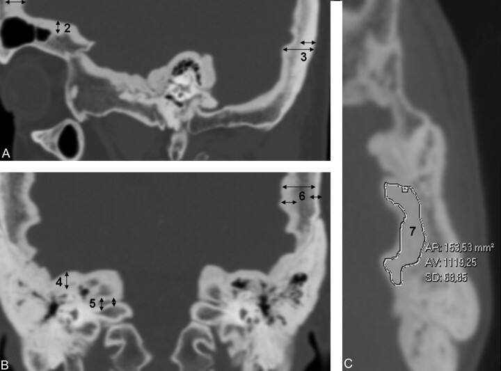
Materials and methods: CT scans of affected individuals within 1 family were analyzed and compared with scans of their unaffected family members and with an age- and sex-matched control group. Linear measurements were performed of the inner table, the medulla, and the outer table of different skull locations, and attenuation (density) measurements of the same regions were recorded. Neuroforamina widths were recorded as well.

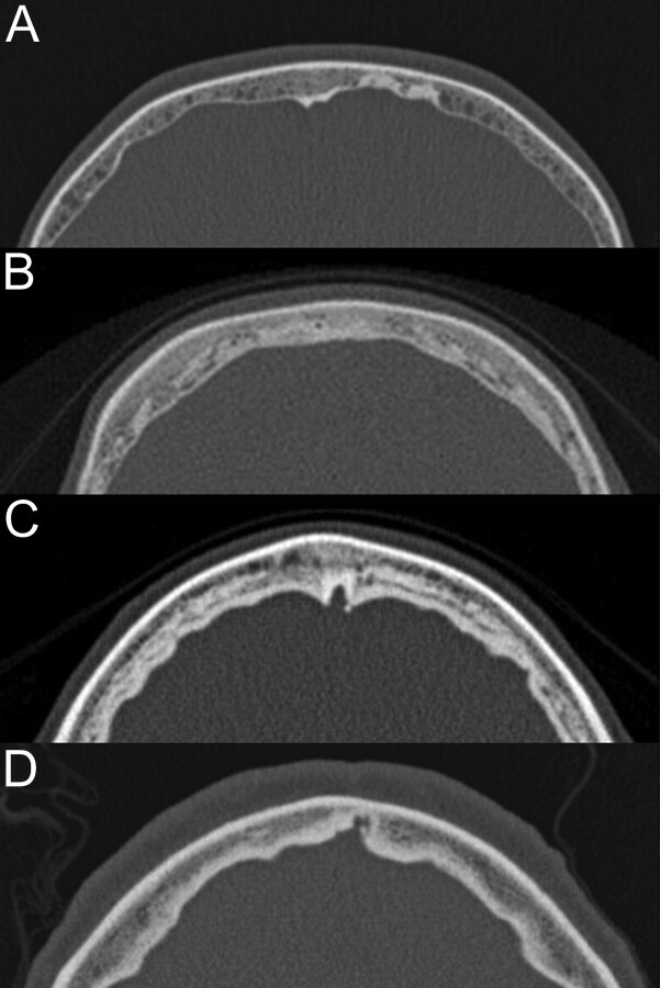
Results: There was significant thickening of the skull in the frontal, parietal, temporal, and occipital regions, which was mainly due to thickening of the inner table of the skull. The attenuation of the deposited hyperostotic bone was lower than normal cortical bone.

Conclusions: HCI is the only genetic bone dysplasia known that is confined to the craniofacial area. The hyperostotic bone is less attenuated than normal cortical bone. The observed radiologic abnormalities explain the possible impairment of the olfactory, optic, trigeminal, facial, and vestibulocochlear nerves.


Figures

Fig 1.
Fig 1.
3D reconstruction of a normal skull base and one of a patient with HCI. The left image is an unaffected individual in whom all neuroforamina can be identified. The right image is an affected individual with thickened calvaria and a bulgy skull base; neuroforamina are hardly visible.
Fig 2.
Fig 2.
Pedigree of the affected kindred. An asterisk is placed if the individual had undergone a CT scan.
Fig 3.
Fig 3.
Simplified examples of different measurements. A and B, linear measurements of skull thickness and neuroforamen width. C, attenuation measurement of freehand region of interest at the inner table of parietal bone. 1, frontal bone measurements; 2, frontal skull base inner table thickness; 3, occipital bone measurements; 4, middle skull base inner table thickness; 5, internal auditory canal measurements (fundus = lateral part, porus = medial part); 6, parietal bone measurements, and 7, parietal inner table attenuation measurement.
Fig 4.
Fig 4.
Evolution of groups 1 and 3 per anatomic region. A, frontal bone; B, parietal bone; C, occipital bone; D, skull base; E, internal auditory canal, and F, miscellaneous. X-axis = age; Y-axis = distance (mm). TI = inner table (tabula interna); TE = outer table (tabula externa).
Fig 5.
Fig 5.
Density profiles of skull transsections in frontal (upper) and frontotemporal (lower) bone (upper) region. X-axis = distance; Y-axis = attenuation (HU). The higher peak correlates with the outer table. Inner table attenuation measurements are found to be lower in affected individuals than outer table attenuation, in contrast to unaffected individuals.
Fig 6.
Fig 6.
Development of abnormalities in the frontal bone, axial images. A, age 9; B, age 16; C, age 23; D, age 68.
Fig 7.
Fig 7.
Temporal bone. A, 9-year-old boy: Hyperostotic superior (asterisk) and inferior lip of the IAC (coronal reconstruction). B, 41-year-old woman: Pinpoint internal auditory canal (arrow, sagittal reconstruction). C, Progressive bilateral narrowing of the IAC in a 16-year-old girl (asterisk, coronal reconstruction). D, Hyperostotic stylomastoid process in a 44-year-old woman (arrow, coronal reconstruction). E–G: Axial images of left temporal bones with extensive hyperostosis in posterior fossa (asterisk) after foramen magnum decompression (double-headed arrow). E, 41-year-old woman with extensive pneumatization progressively substituted by sclerotic bone (F and G). Note the middle ear space is not affected (arrow G).
Fig 8.
Fig 8.
Sphenoid, ethmoid, mandible. A, Axial image of 23-year-old woman: hyperostosis of endosteal greater sphenoid wing, decreased space in temporal lobe area; hyperostotic lamina papyracea and cribriform plate area. Anterior line corresponds with coronal image B (different patient); posterior line corresponds with the coronal image D (same patient). B, Coronal image of 41-year-old woman showing extensive supraorbital pneumatization and inclusion of unaffected crista galli (arrow). C, Axial image of mandibular exostoses in a 68-year-old patient. Note that the inner table of the mandible shows a normal aspect. D, Coronal image (same patient as A) of the greater and lesser sphenoid wings. Note narrowing of the optic canal (upper arrow) and the superior orbital fissure (lower arrow).

References

    1. Manni JJ, Scaf JJ, Huygen PL, et al. . Hyperostosis cranialis interna: a new hereditary syndrome with cranial-nerve entrapment. N Engl J Med 1990; 322: 450– 54 - PubMed
    1. Waterval JJ, Stokroos RJ, Bauer NJ, et al. . Phenotypic manifestations and management of hyperostosis cranialis interna, a hereditary bone dysplasia affecting the calvaria and the skull base. Am J Med Genet A 2010; 152A: 547– 55 - PubMed
    1. Rimoin DL, Cohn D, Krakow D, et al. . The skeletal dysplasias: clinical-molecular correlations. Ann N Y Acad Sci 2007; 1117: 302– 09 - PubMed
    1. Superti-Furga A, Unger S. Nosology and classification of genetic skeletal disorders: 2006 revision. Am J Med Genet A 2007; 143: 1– 18 - PubMed
    1. Skrzat J, Brzegowy P, Walocha J, et al. . Age dependent changes of the diploe in the human skull. Folia Morphol 2004; 63: 67– 70 - PubMed