Review
. 2012 Apr;33(4):586-94.
doi: 10.3174/ajnr.A2535. Epub 2011 Dec 22.

Biologic imaging of head and neck cancer: the present and the future

Affiliations
Review

Biologic imaging of head and neck cancer: the present and the future

A Srinivasan et al. AJNR Am J Neuroradiol. 2012 Apr.

Abstract

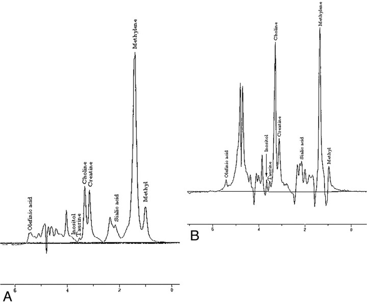
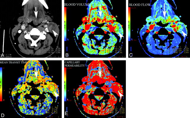
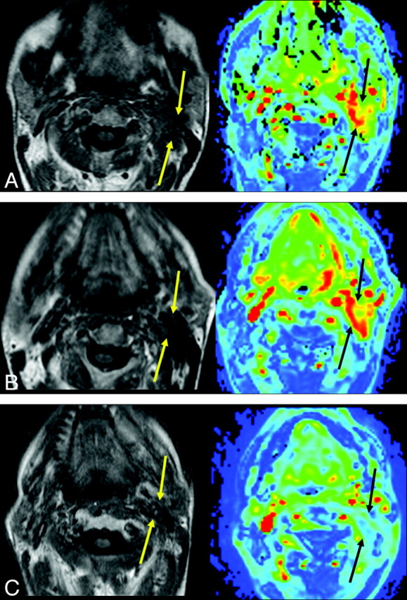
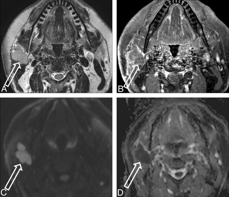
While anatomic imaging (CT and MR imaging) of HNC is focused on diagnosing and/or characterizing the disease, defining its local extent, and evaluating distant spread, accurate assessment of the biologic status of the cancer (cellularity, growth rate, response to nonsurgical chemoradiation therapy, and so forth) can be invaluable for prognostication, planning therapy, and follow-up of lesions after therapy. The combination of anatomic and biologic imaging techniques can thus provide a more comprehensive evaluation of the patient. The purpose of this work was to review the present and future clinical applications of advanced biologic imaging techniques in HNC evaluation and management. As part of the biologic imaging array, we discuss MR spectroscopy, diffusion and perfusion MR imaging, CTP, and FDG-PET scanning and conclude with exciting developments that hold promise in assessment of tumor hypoxia and neoangiogenesis.


Figures

Fig 1.
Fig 1.
Comparison of MR spectra between muscle (A) and squamous cell carcinoma (B) in the head and neck demonstrates elevation of the Cho peak in squamous cell carcinoma. Analysis of other metabolites like amino acids has also been found to be useful in characterization of HNCs.
Fig 2.
Fig 2.
CTP maps generated in a patient with epiglottic cancer and a metastatic necrotic left level II lymph node (A) demonstrate elevated BV (B), elevated BF (C), decreased MTT (D), and increased capillary permeability (E) in both the primary tumor (arrow) and the metastatic node (arrowhead) compared with the normal sternomastoid muscle on the contralateral side.
Fig 3.
Fig 3.
A and B, Axial T1-weighted and BV dynamic contrast-enhanced MR perfusion images in a patient with left neck squamous cell carcinoma demonstrate relatively increased BV (A) on the pretherapy scan, further increase (B) during midtherapy (3 weeks during radiation therapy), and eventual decreases (C) at the end of therapy (6 weeks after completion). The patient had a complete response of tumor to therapy as seen on follow-up CT-PET imaging.
Fig 4.
Fig 4.
Axial T2-weighted (A) and postcontrast T1-weighted (B) images in a 57-year-old patient demonstrate a T2 hyperintense, heterogeneously enhancing lesion arising from the right parotid gland. The lesion is bright on the b = 800 diffusion image (C) and shows restriction of diffusion on the ADC map (D), with the ADC measuring 0.712 × 10⁻³ mm2/s. This was resected with a final pathologic diagnosis of neuroendocrine carcinoma.
Fig 5.
Fig 5.
Pre- and postsurgical MR images in a patient with sinonasal undifferentiated carcinoma within the nasal cavity and ethmoid sinuses. A−D, Presurgical images demonstrate a T2 hypointense (A) enhancing lesion (B) with increased diffusion signal intensity on the b = 800 image (C) and restriction of diffusion (ADC = 0.701 ×10⁻³ mm2/s) on the ADC map (D). Six months after surgery and radiation therapy, the patient presented with epistaxis, prompting re-imaging. E and F, MR imaging shows a small T2 hypointense focus on the left side (E) with intense enhancement (F). G and H, However, the focus is only mildly hyperintense on the b = 800 diffusion image (G) and is not restricting diffusion (ADC = 1.8 × 10⁻³ mm2/s) on the ADC map (H), suggesting that this is probably not tumor recurrence. However, biopsy was still performed and pathology revealed benign granulation tissue. With further support from the literature, it may be possible to avoid biopsies in patients demonstrating ADC values not consistent with tumor recurrence.
Fig 6.
Fig 6.
A, CT scan obtained as part of PET/CT fusion imaging in a patient with orbital squamous cell carcinoma treated with exenteration shows soft tissue at the orbital apex (arrowhead), which did not show any enhancement with contrast administration (not shown). B, Corresponding FDG-PET image shows intense FDG avidity, raising the suspicion for tumor recurrence, which was confirmed on pathology. FDG-PET imaging can be helpful for detection of residual and recurrent malignancies in the head and neck.

References

    1. American Cancer Society. www.cancer.org. Accessed October 28, 2007
    1. Delikatny EJ, Russell P, Hunter JC, et al. . Proton MR and human cervical neoplasia: ex vivo spectroscopy allows distinction of invasive carcinoma of the cervix from carcinoma in situ and other preinvasive lesions. Radiology 1993;188: 791–96 - PubMed
    1. Gill SS, Thomas DG, Van Bruggen N, et al. . Proton MR spectroscopy of intracranial tumours: in vivo and in vitro studies. J Comput Assist Tomogr 1990;14: 497–504 - PubMed
    1. Negendank WG, Brown TR, Evelhoch JL, et al. . Proceedings of a National Cancer Institute workshop: MR spectroscopy and tumor cell biology. Radiology 1992;185: 875–83 - PubMed
    1. Shah GV, Gandhi D, Mukherji SK. Magnetic resonance spectroscopy of head and neck neoplasms. Top Magn Reson Imaging 2004;15: 87–94 - PubMed

MeSH terms

Substances