

A distinct MR imaging phenotype in amyotrophic lateral sclerosis: correlation between T1 magnetization transfer contrast hyperintensity along the corticospinal tract and diffusion tensor imaging analysis
- PMID: 22194369
- PMCID: PMC8050435
- DOI: 10.3174/ajnr.A2855
A distinct MR imaging phenotype in amyotrophic lateral sclerosis: correlation between T1 magnetization transfer contrast hyperintensity along the corticospinal tract and diffusion tensor imaging analysis
Abstract
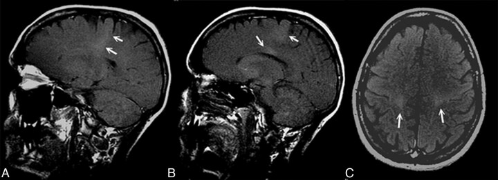
Background and purpose: In the search for a diagnostic marker in ALS, we focused our attention on the hyperintense signal intensity in T1 MTC MR images along the CST, detected in some patients and not found in other patients with ALS and in control subjects. The aim of this study was to investigate the relationship between the hyperintense signal intensity in T1 MTC images and white matter damage. To this purpose, we studied potential heterogeneities in DTI values within our patients by using TBSS without a priori anatomic information.
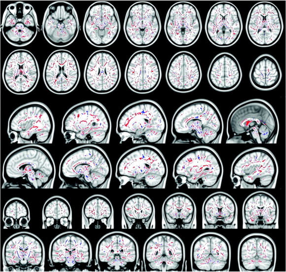
Materials and methods: In 43 patients with ALS and 43 healthy control subjects, the presence or absence of T1 MTC hyperintense signal intensity was evaluated. With a DTI analysis with a TBSS approach, differences in FA distribution between the 2 groups (patients with T1 MTC hyperintense signal intensity and patients without it) compared with each other and with control subjects were investigated.
Results: We found regional differences in white matter FA between patients with T1 MTC hyperintense signal intensity (37.2%) and patients without it. Patients with T1 MTC abnormal signal intensity showed lower FA strictly limited to the motor network and the posterior aspect of the body of the CC without extramotor FA reductions, whereas patients without this sign showed FA reductions in several confluent regions within and outside the CST and in the whole CC.
Conclusions: T1 MTC hyperintense signal intensity in the CST and posterior CC, when present, is specific for ALS and represents, among patients with ALS, a possible distinct phenotype of presentation of the disease with prominent UMN involvement.
Figures
Comment in
-
Corticospinal tract MR signal-intensity pseudonormalization on magnetization transfer contrast imaging: a potential pitfall in the interpretation of the advanced compromise of upper motor neurons in amyotrophic lateral sclerosis.AJNR Am J Neuroradiol. 2012 May;33(5):E79-80. doi: 10.3174/ajnr.A3114. Epub 2012 Mar 29. AJNR Am J Neuroradiol. 2012. PMID: 22460338 Free PMC article. No abstract available.
Similar articles
-
Differential involvement of corticospinal tract (CST) fibers in UMN-predominant ALS patients with or without CST hyperintensity: A diffusion tensor tractography study.Neuroimage Clin. 2017 Feb 22;14:574-579. doi: 10.1016/j.nicl.2017.02.017. eCollection 2017. Neuroimage Clin. 2017. PMID: 28337412 Free PMC article.
-
Detection of corticospinal tract compromise in amyotrophic lateral sclerosis with brain MR imaging: relevance of the T1-weighted spin-echo magnetization transfer contrast sequence.AJNR Am J Neuroradiol. 2004 Oct;25(9):1509-15. AJNR Am J Neuroradiol. 2004. PMID: 15502129 Free PMC article.
-
Widespread microstructural white matter involvement in amyotrophic lateral sclerosis: a whole-brain DTI study.AJNR Am J Neuroradiol. 2012 Jun;33(6):1102-8. doi: 10.3174/ajnr.A2918. Epub 2012 Feb 2. AJNR Am J Neuroradiol. 2012. PMID: 22300932 Free PMC article.
-
[Objective markers for upper motor neuron involvement in amyotrophic lateral sclerosis].Brain Nerve. 2007 Oct;59(10):1053-64. Brain Nerve. 2007. PMID: 17969345 Review. Japanese.
-
The role of diffusion tensor imaging and fractional anisotropy in the evaluation of patients with idiopathic normal pressure hydrocephalus: a literature review.Neurosurg Focus. 2016 Sep;41(3):E12. doi: 10.3171/2016.6.FOCUS16192. Neurosurg Focus. 2016. PMID: 27581308 Review.
Cited by
-
Quantitative assessment of amyotrophic lateral sclerosis with diffusion tensor imaging in 3.0T magnetic resonance.Int J Clin Exp Med. 2015 May 15;8(5):8295-303. eCollection 2015. Int J Clin Exp Med. 2015. PMID: 26221413 Free PMC article.
-
Tract integrity in amyotrophic lateral sclerosis: 6-month evaluation using MR diffusion tensor imaging.BMC Med Imaging. 2019 Feb 22;19(1):19. doi: 10.1186/s12880-019-0319-3. BMC Med Imaging. 2019. PMID: 30795741 Free PMC article.
-
Role of PET and SPECT in the study of amyotrophic lateral sclerosis.Biomed Res Int. 2014;2014:237437. doi: 10.1155/2014/237437. Epub 2014 Apr 10. Biomed Res Int. 2014. PMID: 24818133 Free PMC article. Review.
-
Dementia in motor neuron disease: Reviewing the role of MRI in diagnosis.Dement Neuropsychol. 2015 Oct-Dec;9(4):369-379. doi: 10.1590/1980-57642015DN94000380. Dement Neuropsychol. 2015. PMID: 29213986 Free PMC article. Review.
-
Diffusion tensor MRI of the corpus callosum in amyotrophic lateral sclerosis.J Magn Reson Imaging. 2014 Mar;39(3):641-7. doi: 10.1002/jmri.24218. Epub 2013 Jul 10. J Magn Reson Imaging. 2014. PMID: 23843179 Free PMC article.
References
-
- Davidson CD. Amyotrophic lateral sclerosis: origin and extent of the upper motor neuron lesion. Arch Neurol 1941;46: 1039–56
-
- Chancellor AM, Slattery JM, Fraser H, et al. . The prognosis of adult-onset motor neuron disease: a prospective study based on the Scottish Motor Neuron Disease Register. J Neurol 1993;240: 339–46 - PubMed
-
- Worms PM. The epidemiology of motor neuron diseases: a review of recent studies. J Neurol Sci 2001;191: 3–9 - PubMed
-
- Logroscino G, Traynor BJ, Hardiman O, et al. , for EURALS. Descriptive epidemiology of amyotrophic lateral sclerosis: new evidence and unsolved issues. J Neurol Neurosurg Psychiatry 2008;79: 6–11 - PubMed
Publication types
MeSH terms
LinkOut - more resources
Full Text Sources
Medical
Miscellaneous