Comparative Study
. 2012 Mar;33(3):434-8.
doi: 10.3174/ajnr.A2807. Epub 2011 Dec 22.

CT and ultrasound features of basal cell adenoma of the parotid gland: a report of 22 cases with pathologic correlation

Affiliations
Comparative Study

CT and ultrasound features of basal cell adenoma of the parotid gland: a report of 22 cases with pathologic correlation

L Shi et al. AJNR Am J Neuroradiol. 2012 Mar.

Abstract

Background and purpose: Parotid gland BCA is a rare benign tumor. Only a few studies describing the imaging features of BCA have been published. This study investigated CT and sonography characteristics of BCA of the parotid gland.

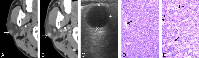
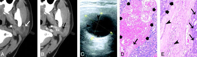
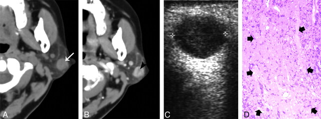
Materials and methods: Demographics of patients with BCA were evaluated, and lesion characteristics of CT (n = 22) and sonography (n = 20) were reviewed. These cases were grouped into 3 types: type 1 tumors, located at the superficial region of superficial lobe of the parotid gland; type 2 tumors, located at the deeper region of superficial lobe; and type 3 tumors, located in the deep lobe. Imaging findings were correlated with pathology.

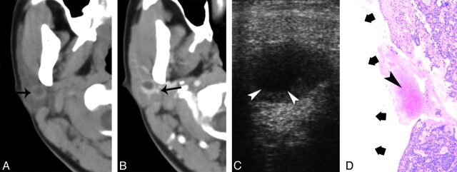
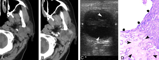
Results: Sixteen patients (73%) were female and 6 (27%) were male. The mean age was 51.5 years (SD 10.2; range 32-73). The size of the tumors was less than 30 mm. The sizes of type 1, type 2, and type 3 tumors were 11.4 ± 3.29 mm, 19.3 ± 5.44 mm, and 26 ± 3.6 mm, respectively. The CT attenuation increase was 64.5 ± 19 HU on contrast CT. The type 1 tumors were solid (11/11), showed homogeneous or slightly heterogeneous enhancement on CT, and were homogeneously or slightly heterogeneously hypoechoic on sonography. Cystic changes tended to occur in type 2 (7/8) or type 3 (2/3) tumors, which showed obvious heterogeneous attenuation on CT and anechoic on sonography.

Conclusions: The BCA tends to be small and shows early intense enhancement. The solid tumor is common in the superficial region of the parotid gland, and cystic lesions occur mostly in the deeper parts of the superficial lobe or in the deep lobe.


Figures

Fig 1.
Fig 1.
A 55-year-old man with type 1 BCA in the right parotid gland A, and B, CT shows a round, well-defined, homogeneously enhancing lesion in the superficial region of the right parotid superficial lobe. The lesion is adjacent to the outer border of the parotid gland (white arrow). C, The tumor shows as well-defined and homogeneously hypoechoic on sonography. D, and E. Photomicrographs of tumor specimen (hematoxylin-eosin stain; ×50 and ×100) show capillaries (black arrows) and uniform tumor cell without hemorrhage, collagen deposition, or cystic change.
Fig 2.
Fig 2.
A 40-year-old woman with type 2 BCA in the left parotid gland. A and B, CT shows a round, ill-defined lesion (white arrow) with a cystic component (black arrow) in the deeper region. C, The lesion shows as heterogeneously hypoechoic, with an anechoic region (arrowheads) on sonography. The superficial border of tumor is irregular and ill defined. D, Photomicrograph (hematoxylin-eosin stain; ×50) demonstrates tumor cystic region with hemorrhage (thick arrows) and collagen deposition (thin arrow). E, Specimen histology (hematoxylin-eosin stain; ×50) shows the tumor tissue (thick arrows), the infiltration of tumor cells into the capsule (arrowhead), and tumor cells in surrounding tissue (thin arrows).
Fig 3.
Fig 3.
A 54-year-old woman with type 1 BCA in the left parotid gland. A, Plain CT shows a round nodule with homogeneous attenuation located at the superficial region of the superficial lobe (white arrow). B, The tumor shows slight heterogeneous enhancement with a small, low attenuation component (black arrowheads) on contrast CT. C, Sonography shows well-defined heterogeneously hypoechoic lesion. D, Correlated to the low attenuation component on the postcontrast CT, a photomicrograph of a tumor section (hematoxylin-eosin stain; ×50) shows a collagen component (thick arrows).
Fig 4.
Fig 4.
A 32-year-old woman with type 2 BCA in the right parotid gland. A, Plain CT shows a round lesion (9 mm) with a cystic area (black arrow) located at the deeper region of the superficial lobe. B, Contrast-enhanced CT shows obvious enhancement of the peripheral tumor rim, whereas the cystic component does not enhance. The tumor is adjacent to the retromandibular vein (black arrow). C, Sonography shows a well-defined, hypoechoic nodule with an anechoic core (white arrowheads). D, Photomicrographs (hematoxylin-eosin stain; ×50) show a hemorrhage (arrowhead) and cystic region (thick arrows) within the tumor.
Fig 5.
Fig 5.
A 73-year-old man with type 3 BCA in the left parotid gland. A, Plain CT shows a round lesion with cystic area (black arrow). B, The lesion displays obvious heterogeneous enhancement on contrast CT scan. C, Sonography shows a well-defined hypoechoic tumor with an anechoic region (white arrowheads). D, Photomicrographs of tumor specimen (hematoxylin-eosin stain; ×50) show collagen deposition (arrow head) and tumor cystic component (thick arrows).

References

    1. Lee YY, Wong KT, King AD, et al. . Imaging of salivary gland tumours. Eur J Radiol 2008; 66: 419– 36 - PubMed
    1. Nagao K, Matsuzaki O, Saiga H, et al. . Histopathologic studies of basal cell adenoma of the parotid gland. Cancer 1982; 50: 736– 45 - PubMed
    1. Kawata R, Yoshimura K, Lee K, et al. . Basal cell adenoma of the parotid gland: a clinicopathological study of nine cases—basal cell adenoma versus pleomorphic adenoma and Warthin's tumor. Eur Arch Otorhinolaryngol 2010; 267: 779– 83 - PubMed
    1. Kleinsasser O, Klein HJ. Basal cell adenoma of the salivary glands [in German]. Arch Klin Exp Ohren Nasen Kehlkopfheilkd 1967; 189: 302– 16 - PubMed
    1. Chawla AJ, Tan TY, Tan GJ. Basal cell adenoma of the parotid gland: CT scan features. Eur J Radiol 2006; 58: 260– 65 - PubMed

Publication types