

When should I do dynamic CT myelography? Predicting fast spinal CSF leaks in patients with spontaneous intracranial hypotension
- PMID: 22194380
- PMCID: PMC8050426
- DOI: 10.3174/ajnr.A2849
When should I do dynamic CT myelography? Predicting fast spinal CSF leaks in patients with spontaneous intracranial hypotension
Abstract
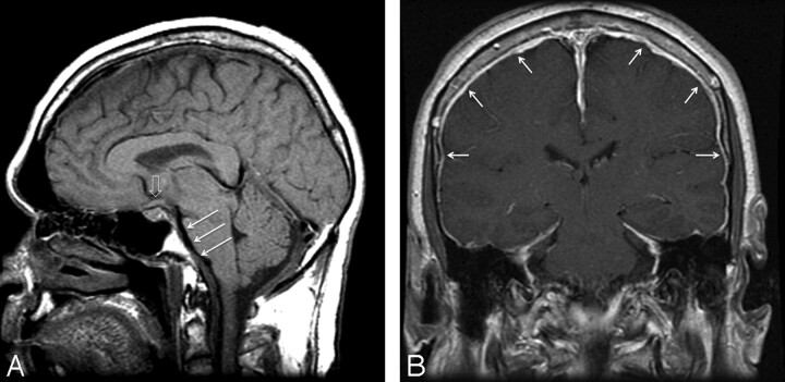
Background and purpose: Some patients with SIH have fast CSF leaks requiring dynamic CTM for localization; however, patients generally undergo conventional CTM before a dynamic study. Our aim was to determine whether findings on head MR imaging, spine MR imaging, or opening pressure measurements can predict fast spinal CSF leaks.
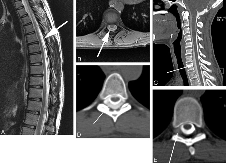
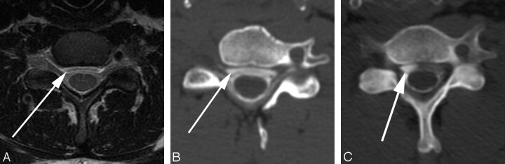
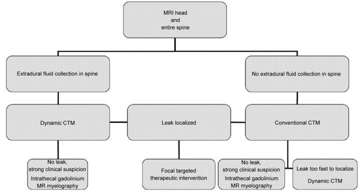
Materials and methods: A retrospective review was performed on 151 consecutive patients referred for CTM to evaluate for spinal CSF leak. Head MR imaging was evaluated for diffuse dural enhancement and "brain sag," and spine MR imaging for presence of an extradural fluid collection. The opening pressure was recorded. The CTM was scored as no leak, slow leak localized on conventional CTM, or fast leak that required dynamic CTM.
Results: Fast CSF leaks were identified in 32 (21%), slow leaks in 36 (24%), and no leak in 83 (55%) of 151 patients on initial CTM. There was significant association between spinal extra-arachnoid fluid on MR imaging and the presence of a fast leak (sensitivity 85%, specificity 79%, P < .0001). There was not significant association between fast leak and findings on head MR imaging (P = .27) or opening pressure (P = .30).
Conclusions: If all patients with spinal extra-arachnoid CSF on MR imaging had been sent directly to dynamic CTM, repeat myelography would have been avoided in most patients with fast leaks (23 of 27; 85%). However, a minority of patients with slow or no leaks would have been converted from conventional to dynamic CTM (16 of 77; 21%). Spinal MR imaging is helpful in premyelographic evaluation of SIH.
Figures
References
-
- Mokri B. Spontaneous intracranial hypotension. Curr Neurol Neurosci Rep 2001;1: 109–17 - PubMed
-
- Thielen KR, Sillery J, Morris JM, et al. . Ultrafast dynamic CT myelography for the precise identification of high-flow cerebrospinal fluid leaks caused by spiculated osteophytes of the spine. Annual Meeting of the American Society of Neuroradiology, Seattle, Washington, June 6, 2011.
-
- Mokri B. Spontaneous low cerebrospinal pressure/volume headaches. Curr Neurol Neurosci Rep 2004;4: 117–24 - PubMed
-
- Mokri B, Krueger BR, Miller GM, et al. . Meningeal gadolinium enhancemnet in low pressure headaches [abstract]. Ann Neurol 1991;30: 294–95
MeSH terms
LinkOut - more resources
Full Text Sources
Medical