. 2012 Feb 1;72(3):810-20.
doi: 10.1158/0008-5472.CAN-11-1052. Epub 2011 Dec 22.

SMYD3 promotes cancer invasion by epigenetic upregulation of the metalloproteinase MMP-9

Affiliations

SMYD3 promotes cancer invasion by epigenetic upregulation of the metalloproteinase MMP-9

Alicia M Cock-Rada et al. Cancer Res. .

Abstract

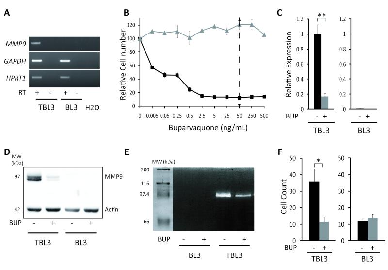
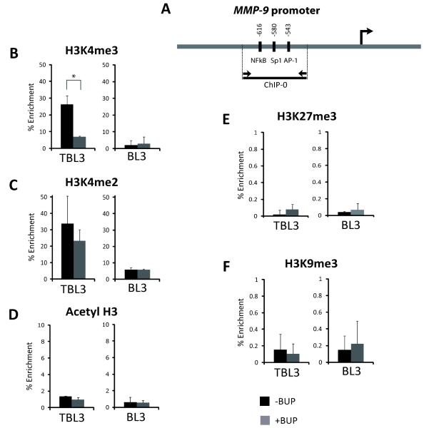
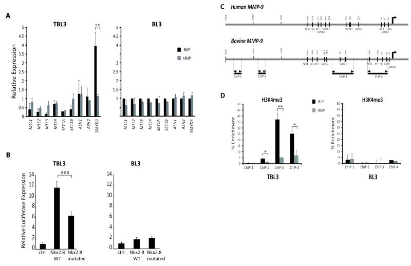
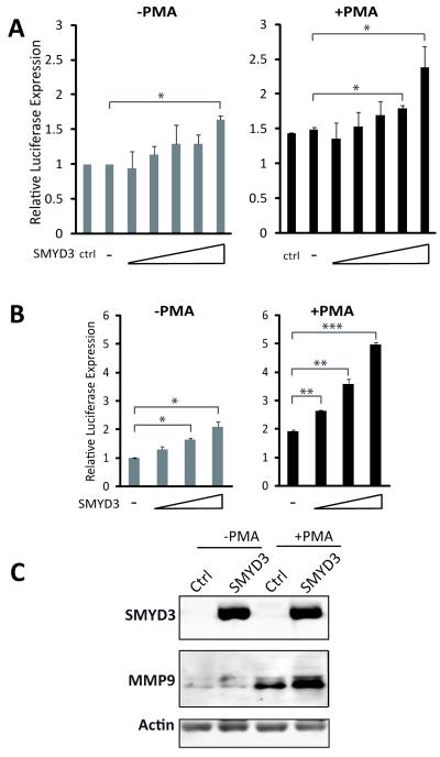
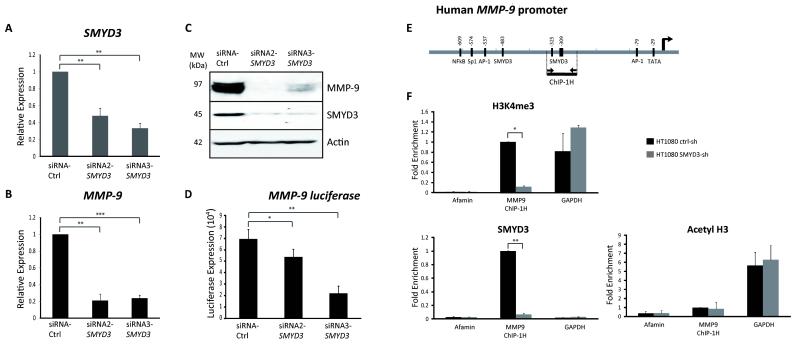
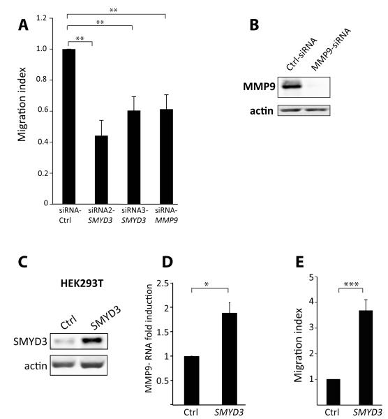
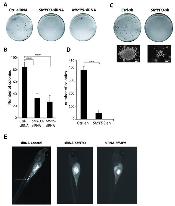
Upregulation of the matrix metalloproteinase (MMP)-9 plays a central role in tumor progression and metastasis by stimulating cell migration, tumor invasion, and angiogenesis. To gain insights into MMP-9 expression, we investigated its epigenetic control in a reversible model of cancer that is initiated by infection with intracellular Theileria parasites. Gene induction by parasite infection was associated with trimethylation of histone H3K4 (H3K4me3) at the MMP-9 promoter. Notably, we found that the H3K4 methyltransferase SMYD3 was the only histone methyltransferase upregulated upon infection. SMYD3 is overexpressed in many types of cancer cells, but its contributions to malignant pathophysiology are unclear. We found that overexpression of SMYD3 was sufficient to induce MMP-9 expression in transformed leukocytes and fibrosarcoma cells and that proinflammatory phorbol esters further enhanced this effect. Furthermore, SMYD3 was sufficient to increase cell migration associated with MMP-9 expression. In contrast, RNA interference-mediated knockdown of SMYD3 decreased H3K4me3 modification of the MMP-9 promoter, reduced MMP-9 expression, and reduced tumor cell proliferation. Furthermore, SMYD3 knockdown also reduced cellular invasion in a zebrafish xenograft model of cancer. Together, our results define SMYD3 as an important new regulator of MMP-9 transcription, and they provide a molecular link between SMYD3 overexpression and metastatic cancer progression.


Figures

Figure 1
Figure 1. Inducible and reversible MMP-9 expression upon Theileria-transformation
A) MMP-9 expression in infected (TBL3) and non-infected (BL3) cells by semiquantitative PCR analysis. mRNAs were incubated with or without Reverse Transcriptase (RT). Two housekeeping genes, GAPDH and HPRT1, were used as controls. B) The relative number of TBL3 (squares) and BL3 (triangles) cells incubated with increasing concentrations of Buparvaquone assessed using the XTT assay. The arrow represents the concentration used for all subsequent experiments. C) The reversibility of MMP-9 expression was assessed by qPCR analysis. Cells were treated with the drug Buparvaquone (BUP) for 64 hours. β-actin mRNA was used for normalization. D) Western blot analysis of MMP-9 protein expression in TBL3/BL3 cells with/without Buparvaquone treatment for 64 hours. Bovine β-actin was used as a control. MW is the molecular weight marker (kDa). E) Gelatin zymography to analyse MMP-9 protein activity in supernatants from BL3 and infected TBL3 cells with/without Buparvaquone treatment. F) Invasion capacity tested using a modified Boyden chamber assay with/without Buparvaquone (BUP). Results show the number of invasive cells counted under the microscope. All results represent the average of three independent experiments (mean+/−sd) *p<0.05, **p<0.01
Figure 2
Figure 2. Chromatin analysis of bovine MMP-9 promoter upon Theileria infection
A) Primers (ChIP-0) were designed to amplify by qPCR a sequence surrounding the NF-κB and AP-1 binding sites in the distal bovine MMP-9 promoter. The right arrow marks the transcription start site. (B-F) BL3 and Theileria-infected TBL3 cells were incubated with/without Buparvaquone (BUP) for 64 hours. Chromatin Immunoprecipitation analysis with antibodies recognizing (B) H3K4me3, (C) H3K4me2, (D) Acetyl-H3, (E) H3K27me3 and (F) H3K9me3 marks. Error bars are representative of two independent experiments. *p<0.05
Figure 3
Figure 3. SMYD3 methyltransferase is upregulated in Theileria-infected cells
A) RT-qPCR analysis of the expression levels of H3K4-methyltransferases using RNA from Theileria-infected TBL3 or BL3 cells, with/without Buparvaquone treatment (BUP) for 64 hours. Beta-actin mRNA was used for normalization. B) SMYD3 activity measured by a dual-luciferase assay in TBL3/BL3 cells transfected with a SMYD3 reporter (Wild-type Nkx2.8 WT promoter-Luciferase vector), compared to a control vector (Ctrl). Promoter activity was reduced when the SMYD3 binding site was mutated (Nkx2.8 mutated). Transfection efficiency was normalized with co-transfection of a Renilla-encoding plasmid. C) Schematic of the human and bovine MMP-9 promoters highlighting potential SMYD3 binding sites. Oligonucleotides to amplify fragments of the bovine MMP-9 promoter: ChIP-1 (−4.7 kb upstream), ChIP-2 (−1.7 Kb), ChIP-3 (three putative SMYD3 binding sites) and ChIP-4 (proximal AP-1 site). The right arrows mark the transcription start site. D) TBL3/BL3 cells were treated with/without Buparvaquone for 64 hours and Chromatin immunoprecipitation was performed with an antibody recognizing H3K4me3. qPCR analysis was performed with the primer sets mentioned above. Error bars represent three independent experiments. *p<0.05, **p<0.01, ***p<0.001
Figure 4
Figure 4. SMYD3 induces MMP-9 expression in human fibrosarcoma cells
A) The luciferase activity of an integrated MMP-9 reporter construct assessed in HT1080-2.2 cells, with increasing amounts of exogenous SMYD3 expression, with/without PMA treatment. The effects of SMYD3 transfection were compared to non-transfected controls (ctrl) and vector-transfected mock (-)** cells. B) Luciferase assays showing expression of an MMP-9-luciferase construct transfected into HT1080 cells, with increasing amounts of ectopic SMYD3 expression, with/without PMA treatment compared to vector-transfected mock controls (-). (C) Western blot analysis showing the expression of exogenous-transfected SMYD3 and upregulation of endogenous MMP-9 protein in HT1080 cells transfected with SMYD3 or control vectors (Ctrl), with/without PMA treatment. *p<0.05, **p<0.01, ***p<0.001
Figure 5
Figure 5. SMYD3 knockdown causes epigenetic repression of MMP-9 in human fibrosarcoma cells
A and B) HT1080 cells were transfected with two different siRNA oligonucleotides against SMYD3 or control followed by RTqPCR analysis of SMYD3 (A) and MMP-9 expression (B), 48 hours after transfection. GAPDH mRNA was used for normalization. C) Western analysis in HT1080 cells transfected with two different siRNA oligonucleotides against SMYD3 or control siRNA, showing SMYD3 and MMP-9 proteins. β-actin was used as control. MW=molecular weight marker. D) Luciferase activity in HT1080-2.2 cells with an integrated MMP-9 promoter-Luciferase reporter, following transfection with siRNA oligonucleotides against SMYD3 or control siRNA (Ctrl). E) Amplicon surrounding putative SMYD3 binding sites (ChIP-1H) in the human MMP-9 promoter. The right arrow marks the transcription start site. F) Chromatin Immunoprecipitation was performed with antibodies recognizing SMYD3, H3K4me3 or Acetylated-H3. GAPDH promoter is a positive control for the histone marks, and the Afamin promoter a negative control. *p<0.05, **p<0.01, ***p<0.001
Figure 6
Figure 6. SMYD3 modulation affects cell migration of human cell lines
A) HT1080 cells were transfected with two different siRNA oligonucleotides against SMYD3 or against MMP-9 or control siRNA (Ctrl), followed by analysis of cell migration on collagen-coated filters. Results are shown with respect to controls and error bars represent three independent experiments. B) Western blot analysis showing the efficiency of siRNA MMP-9 knockdown in HT1080 cells. Beta-actin was used as control. C) HEK293T cells were transfected with SMYD3-expressing vector or control vector (Ctrl). Exogenous SMYD3 protein levels were analysed by Western blot analysis and Beta-actin was used as control. D) SMYD3 transfection into HEK293T cells caused an upregulation of endogenous MMP-9 levels as assessed by RT-qPCR analysis. E) SMYD3 over-expression in HEK293T cells caused a significant increase in cell migration, compared to Control (Ctrl) transfected cells. Error bars represent three independent experiments. *p<0.05, **p<0.01, ***p<0.001
Figure 7
Figure 7. SMYD3 knockdown reduces tumor cell growth in vitro and invasion in vivo
A) HT1080 cells were transfected with siRNA oligonucleotides against SMYD3 or MMP-9 or control siRNA (Ctrl) followed by soft-agar colony formation assay. Representative wells for each condition are shown. B) Average colony numbers for three independent experiments comparing Control, SMYD3 or MMP-9 siRNA transfected cells. C-D) Colony formation assays (plates, photo micrographs of individual colonies, and colony numbers) as above, were performed using stable clones using shRNA against SMYD3 or control (Ctrl-sh). E) Tumor dissemination in zebrafish embryos: HT1080 cells transfected with control (Ctrl-siRNA), SMYD3-siRNA or MMP9-siRNA were injected into the embryos. Images show representative examples of xenografted embryos. Disseminated tumor cells in the yolk are marked with an arrow. *p<0.05, **p<0.01, ***p<0.001

Comment in

References

    1. Fearon ER, Vogelstein B. A genetic model for colorectal tumorigenesis. Cell. 1990;61:759–67. - PubMed
    1. Esteller M. Cancer epigenomics: DNA methylomes and histone-modification maps. Nat Rev Genet. 2007;8:286–98. - PubMed
    1. Chaussepied M, Langsley G. Theileria transformation of bovine leukocytes: a parasite model for the study of lymphoproliferation. Res Immunol. 1996;147:127–38. - PubMed
    1. Dobbelaere D, Heussler V. Transformation of leukocytes by Theileria parva and T. annulata. Annu Rev Microbiol. 1999;53:1–42. - PubMed
    1. Lizundia R, Chaussepied M, Huerre M, Werling D, Di Santo JP, Langsley G. c-Jun NH2-terminal kinase/c-Jun signaling promotes survival and metastasis of B lymphocytes transformed by Theileria. Cancer Res. 2006;66:6105–10. - PubMed

Publication types

MeSH terms

LinkOut - more resources