. 2011 Dec 23;334(6063):1723-7.
doi: 10.1126/science.1209740.

The ribosome modulates nascent protein folding

Affiliations

The ribosome modulates nascent protein folding

Christian M Kaiser et al. Science. .

Abstract

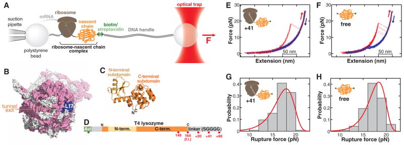
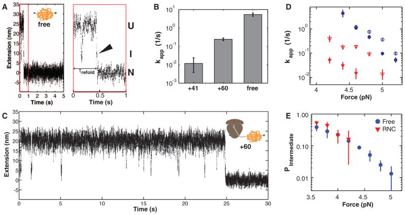
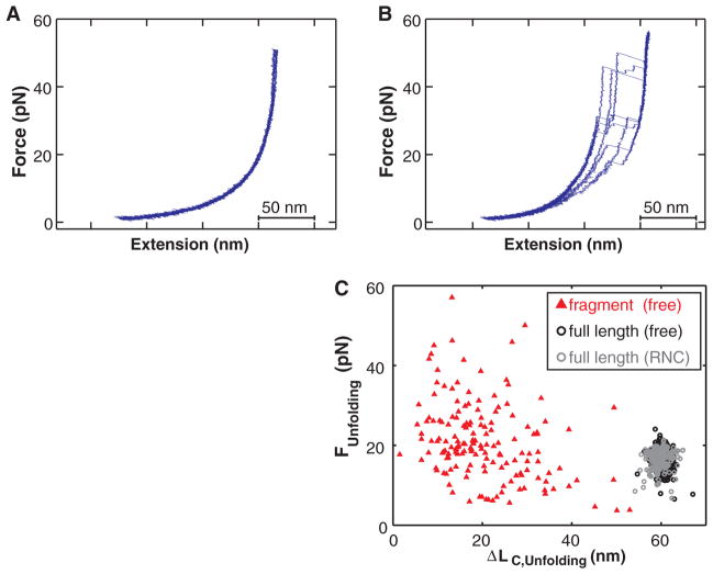
Proteins are synthesized by the ribosome and generally must fold to become functionally active. Although it is commonly assumed that the ribosome affects the folding process, this idea has been extremely difficult to demonstrate. We have developed an experimental system to investigate the folding of single ribosome-bound stalled nascent polypeptides with optical tweezers. In T4 lysozyme, synthesized in a reconstituted in vitro translation system, the ribosome slows the formation of stable tertiary interactions and the attainment of the native state relative to the free protein. Incomplete T4 lysozyme polypeptides misfold and aggregate when free in solution, but they remain folding-competent near the ribosomal surface. Altogether, our results suggest that the ribosome not only decodes the genetic information and synthesizes polypeptides, but also promotes efficient de novo attainment of the native state.


Figures

Fig. 1
Fig. 1
(A) Schematic of the molecular assembly for optical-tweezers experiments. Force can be applied to the nascent polypeptide by moving the optical trap relative to the pipette. (B) Surface representation of the ribosome [Protein Data Bank identification numbers (PDB IDs): 2aw4 and 2avy] showing the opening of the ribosomal exit tunnel in the large subunit and the location of ribosomal protein L17, serving as the attachment site in the optical-tweezers experiments. Ribosomal RNA, pink; ribosomal proteins, white; L17, blue. The small subunit is shown in a semitransparent rendering. (C) Cartoon diagram of T4 lysozyme (PDB ID: 4lzm). The N-terminal subdomain (light orange) is composed of residues 13 to 59; the C-terminal subdomain (dark orange) comprises residues 1 to 12 and 60 to 164. (D) Primary structure diagram of the protein construct translated for optical-tweezers experiments (32). Red spheres indicate stalling positions along the sequence. f.l., full length. (E and F) Force-extension traces of T4 lysozyme unfolding (red) and refolding (blue) on the ribosome (41–amino acid linker) (E) and free in solution (F). The protein unfolds in one cooperative transition near 17 pN. (G and H) Rupture-force histograms (gray bars) for unfolding of the ribosome-bound (G) and free (H) protein. Red lines, rupture force distribution reconstructed from the force-dependent lifetimes (fig. S5).
Fig. 2
Fig. 2
(A) Example extension-versus-time trace of T4 lysozyme refolding at constant force (5 pN). Before transitioning to the native state (N) at ~0.5 s, the protein visits a folding intermediate (I). Enlargement of the first second of the trace (red dashed box) reveals that the intermediate is visited immediately before folding to the native state (arrowhead), demonstrating that the intermediate is on-pathway. U, unfolded state. (B) Apparent refolding rates for ribosome-bound T4 lysozyme with 41–amino acid (+41) and 60–amino acid (+60) linkers and for the free protein (free). Error bars: 95% CIs. (C) Example trace of ribosome-bound protein (60–amino acid linker) re-folding at 5 pN. (D) Apparent refolding rates of free protein (blue circles) and +60 linkers (red triangles) at 150 mM (filled symbols) and 500 mM (open symbols) KCl. Increasing the salt concentration mitigates the effect of the ribosome on the refolding rate. (E) Population of the folding intermediate before refolding. The unfolded and intermediate states are equally populated (P = 0.5) at a force of ~3.6 pN, both in the free and the ribosome-bound protein.
Fig. 3
Fig. 3
(A) Kinetic rates along the T4 lysozyme folding pathway. The rate of the final, irreversible step (kI-N) along the folding pathway is significantly slower for the ribosome-bound protein (60–amino acid linker). (B) Distance changes upon unfolding (NU, open symbols) and refolding (UN, filled symbols) at various forces. At low forces, the distance is shorter than expected from a WLC model (gray line), indicating a partial compaction of the polypeptide that does not resist forces above 4 pN. The compact structure is stabilized in the ribosome-bound protein. Error bars: 95% CIs. (C) Schematic folding energy landscapes of free and ribosome-bound T4 lysozyme. The height of the barrier from I to N is affected by the ribosome, resulting in a decrease in kI-N.
Fig. 4
Fig. 4
(A) Force-extension curves of a T4 lysozyme (164 amino acids) with a 20–amino acid C-terminal extension bound to the ribosome (~150 amino acids outside the ribosomal tunnel), exhibiting no defined unfolding transitions. (B) A free protein fragment of 149 amino acids misfolds into a heterogeneous ensemble of structures that unfold over a wide rage of forces. (C) Comparison of unfolding events recorded for the free protein and the 149–amino acid fragment. The full-length protein unfolds within a narrow, stochastic range of forces and extension changes, whereas the unfolding transitions of the isolated fragment are highly heterogeneous.

References

    1. Anfinsen CB. Science. 1973;181:223. - PubMed
    1. Kubelka J, Chiu TK, Davies DR, Eaton WA, Hofrichter J. J Mol Biol. 2006;359:546. - PubMed
    1. Plaxco KW, Simons KT, Baker D. J Mol Biol. 1998;277:985. - PubMed
    1. Brockwell DJ, Radford SE. Curr Opin Struct Biol. 2007;17:30. - PMC - PubMed
    1. Udgaonkar JB. Annu Rev Biophys. 2008;37:489. - PubMed

Publication types