

Identification of mono- and disulfated N-acetyl-lactosaminyl Oligosaccharide structures as epitopes specifically recognized by humanized monoclonal antibody HMOCC-1 raised against ovarian cancer
- PMID: 22194598
- PMCID: PMC3307324
- DOI: 10.1074/jbc.M111.305334
Identification of mono- and disulfated N-acetyl-lactosaminyl Oligosaccharide structures as epitopes specifically recognized by humanized monoclonal antibody HMOCC-1 raised against ovarian cancer
Abstract
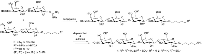
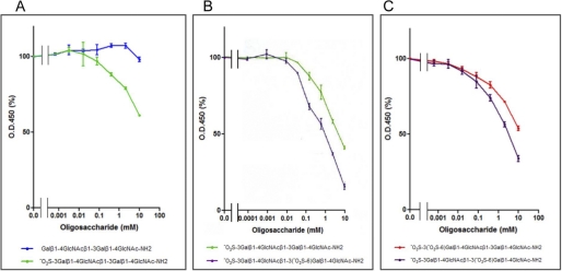
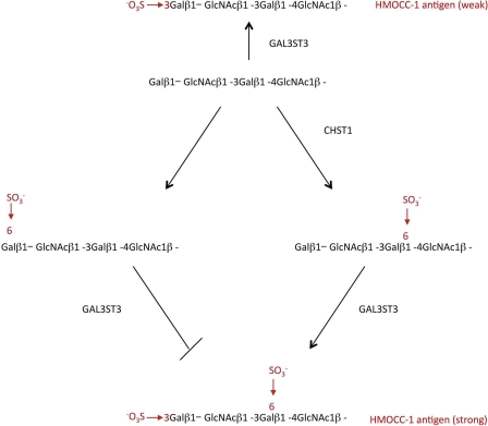
A humanized monoclonal antibody raised against human ovarian cancer RMG-I cells and designated as HMOCC-1 (Suzuki, N., Aoki, D., Tamada, Y., Susumu, N., Orikawa, K., Tsukazaki, K., Sakayori, M., Suzuki, A., Fukuchi, T., Mukai, M., Kojima-Aikawa, K., Ishida, I., and Nozawa, S. (2004) Gynecol. Oncol. 95, 290-298) was characterized for its carbohydrate epitope structure. Specifically, a series of co-transfections was performed using mammalian expression vectors encoding specific glycosyltransferases and sulfotransferases. These experiments identified one sulfotransferase, GAL3ST3, and one glycosyltransferase, B3GNT7, as required for HMOCC-1 antigen formation. They also suggested that the sulfotransferase CHST1 regulates the abundance and intensity of HMOCC-1 antigen. When HEK293T cells were co-transfected with GAL3ST3 and B3GNT7 expression vectors, transfected cells weakly expressed HMOCC-1 antigen. When cells were first co-transfected with GAL3ST3 and B3GNT7 and then with CHST1, the resulting cells strongly expressed HMOCC-1 antigen. However, when cells were transfected with a mixture of GAL3ST3 and CHST1 before or after transfection with B3GNT7, the number of antigen-positive cells decreased relative to the number seen with only GAL3ST3 and B3GNT7, suggesting that CHST1 plays a regulatory role in HMOCC-1 antigen formation. Because these results predicted that HMOCC-1 antigens are SO(3) → 3Galβ1 → 4GlcNAcβ1 → 3(±SO(3) → 6)Galβ1 → 4GlcNAc, we chemically synthesized mono- and disulfated and unsulfated oligosaccharides. Immunoassays using these oligosaccharides as inhibitors showed the strongest activity by disulfated tetrasaccharide, weak but positive activity by monosulfated tetrasaccharide at the terminal galactose, and no activity by nonsulfated tetrasaccharides. These results establish the HMOCC-1 epitope, which should serve as a useful reagent to further characterize ovarian cancer.
Figures
Similar articles
-
HMOCC-1, a human monoclonal antibody that inhibits adhesion of ovarian cancer cells to human mesothelial cells.Gynecol Oncol. 2004 Nov;95(2):290-8. doi: 10.1016/j.ygyno.2004.06.024. Gynecol Oncol. 2004. PMID: 15491748
-
Human monoclonal antibody for ovarian clear cell carcinoma-2, a human monoclonal antibody with antitumor activity against ovarian cancer cells that recognizes CA125-like antigen.Int J Gynecol Cancer. 2008 Sep-Oct;18(5):996-1006. doi: 10.1111/j.1525-1438.2007.01147.x. Epub 2007 Nov 20. Int J Gynecol Cancer. 2008. PMID: 18028379
-
The binding specificity of the marker antibodies Tra-1-60 and Tra-1-81 reveals a novel pluripotency-associated type 1 lactosamine epitope.Glycobiology. 2011 Sep;21(9):1125-30. doi: 10.1093/glycob/cwq209. Epub 2010 Dec 15. Glycobiology. 2011. PMID: 21159783 Free PMC article.
-
Molecular mimicry of host structures by lipooligosaccharides of Neisseria meningitidis: characterization of sialylated and nonsialylated lacto-N-neotetraose (Galbeta1-4GlcNAcbeta1-3Galbeta1-4Glc) structures in lipooligosaccharides using monoclonal antibodies and specific lectins.Adv Exp Med Biol. 2001;491:525-42. doi: 10.1007/978-1-4615-1267-7_35. Adv Exp Med Biol. 2001. PMID: 14533820 Review.
-
Alteration of oligosaccharide biosynthesis by genetic manipulation of glycosyltransferases.Ann N Y Acad Sci. 1994 Nov 30;745:331-5. doi: 10.1111/j.1749-6632.1994.tb44386.x. Ann N Y Acad Sci. 1994. PMID: 7832520 Review.
Cited by
-
KSGal6ST generates galactose-6-O-sulfate in high endothelial venules but does not contribute to L-selectin-dependent lymphocyte homing.Glycobiology. 2013 Mar;23(3):381-94. doi: 10.1093/glycob/cws166. Epub 2012 Dec 18. Glycobiology. 2013. PMID: 23254996 Free PMC article.
-
Determination of carbohydrate structure recognized by prostate-specific F77 monoclonal antibody through expression analysis of glycosyltransferase genes.J Biol Chem. 2014 Jun 6;289(23):16478-86. doi: 10.1074/jbc.M114.559047. Epub 2014 Apr 21. J Biol Chem. 2014. PMID: 24753248 Free PMC article.
-
Characterization of H type 1 and type 1 N-acetyllactosamine glycan epitopes on ovarian cancer specifically recognized by the anti-glycan monoclonal antibody mAb-A4.J Biol Chem. 2017 Apr 14;292(15):6163-6176. doi: 10.1074/jbc.M116.768887. Epub 2017 Feb 6. J Biol Chem. 2017. PMID: 28167527 Free PMC article.
-
Priming mass spectrometry-based sulfoglycomic mapping for identification of terminal sulfated lacdiNAc glycotope.Glycoconj J. 2013 Feb;30(2):183-94. doi: 10.1007/s10719-012-9396-z. Epub 2012 Jun 1. Glycoconj J. 2013. PMID: 22653491
-
Efficient Mapping of Sulfated Glycotopes by Negative Ion Mode nanoLC-MS/MS-Based Sulfoglycomic Analysis of Permethylated Glycans.Anal Chem. 2015 Jun 16;87(12):6380-8. doi: 10.1021/acs.analchem.5b01409. Epub 2015 Jun 5. Anal Chem. 2015. PMID: 26016788 Free PMC article.
References
-
- Hakomori S. (1989) Aberrant glycosylation in tumors and tumor-associated carbohydrate antigens. Adv. Cancer Res. 52, 257–331 - PubMed
-
- Fukuda M. (1996) Possible roles of tumor-associated carbohydrate antigens. Cancer Res. 56, 2237–2244 - PubMed
-
- Kannagi R. (2000) Monoclonal anti-glycosphingolipid antibodies. Methods Enzymol. 312, 160–179 - PubMed
-
- Nakamori S., Kameyama M., Imaoka S., Furukawa H., Ishikawa O., Sasaki Y., Kabuto T., Iwanaga T., Matsushita Y., Irimura T. (1993) Increased expression of sialyl Lewisx antigen correlates with poor survival in patients with colorectal carcinoma. Clinicopathological and immunohistochemical study. Cancer Res. 53, 3632–3637 - PubMed
-
- Kannagi R., Fukushi Y., Tachikawa T., Noda A., Shin S., Shigeta K., Hiraiwa N., Fukuda Y., Inamoto T., Hakomori S. (1986) Quantitative and qualitative characterization of human cancer-associated serum glycoprotein antigens expressing fucosyl or sialyl-fucosyl type 2 chain polylactosamine. Cancer Res. 46, 2619–2626 - PubMed
Publication types
MeSH terms
Substances
Grants and funding
LinkOut - more resources
Full Text Sources
Medical
Research Materials
