. 2012 Feb 10;287(7):4790-9.
doi: 10.1074/jbc.M111.302174. Epub 2011 Dec 21.

Competition between sumoylation and ubiquitination of serine hydroxymethyltransferase 1 determines its nuclear localization and its accumulation in the nucleus

Affiliations

Competition between sumoylation and ubiquitination of serine hydroxymethyltransferase 1 determines its nuclear localization and its accumulation in the nucleus

Donald D Anderson et al. J Biol Chem. .

Abstract

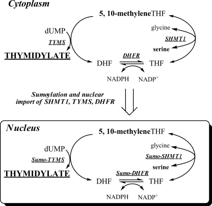
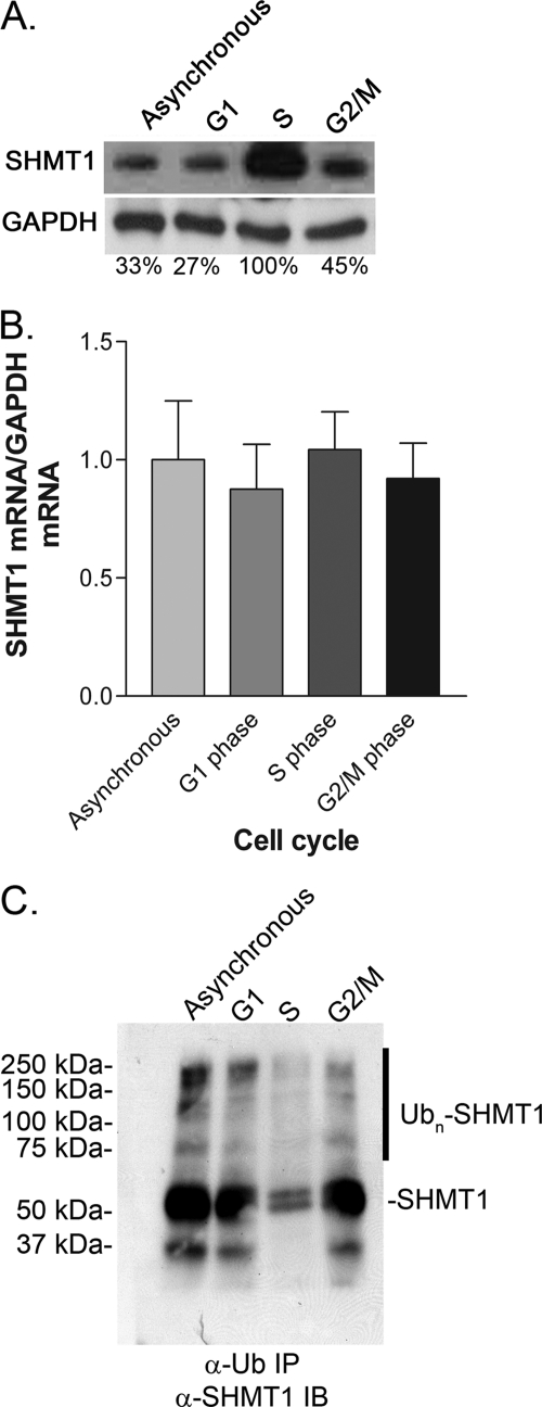
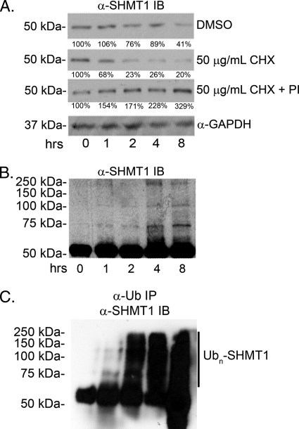
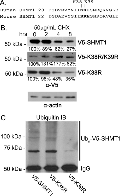
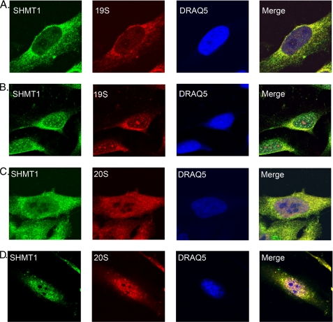
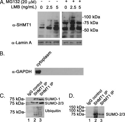
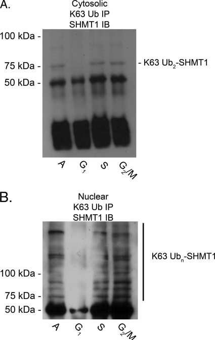
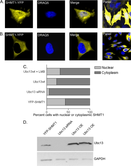
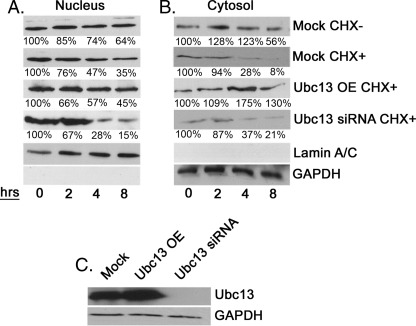
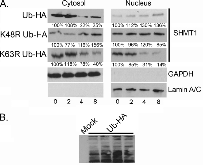
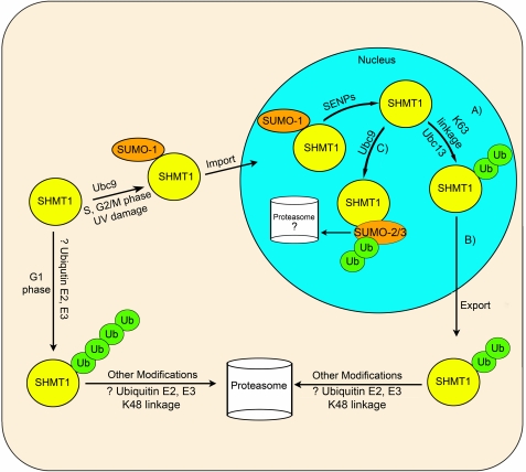
Serine hydroxymethyltransferase 1 (SHMT1) expression limits rates of de novo dTMP synthesis in the nucleus. Here we report that SHMT1 is ubiquitinated at the small ubiquitin-like modifier (SUMO) consensus motif and that ubiquitination at that site is required for SHMT1 degradation. SHMT1 protein levels are cell cycle-regulated, and Ub-SHMT1 levels are lowest at S phase when SHMT1 undergoes SUMO modification and nuclear transport. Mutation of the SUMO consensus motif increases SHMT1 stability. SHMT1 interacts with components of the proteasome in both the nucleus and cytoplasm, indicating that degradation occurs in both compartments. Ubc13-mediated ubiquitination is required for SHMT1 nuclear export and increases stability of SHMT1 within the nucleus, whereas Ubc9-mediated modification with Sumo2/3 is involved in nuclear degradation. These data demonstrate that SUMO and ubiquitin modification of SHMT1 occurs on the same lysine residue and determine the localization and accumulation of SHMT1 in the nucleus.


Figures

FIGURE 1.
FIGURE 1.
Folate-dependent de novo thymidylate biosynthesis. The de novo thymidylate biosynthesis pathway is comprised of SHMT1, thymidylate synthase (TYMS), and DHFR. During S phase, these enzymes are SUMOylated by Ubc9, which serves as a signal for nuclear import.
FIGURE 2.
FIGURE 2.
SHMT1 protein levels change with the cell cycle. HeLa cells were cell cycle-blocked by 24-h treatment with lovastatin (30 μm) for G1 phase, hydroxyurea (1 mm) for S phase, and nocodazole (100 ng/ml) for G2/M phase. A, SHMT1 protein levels were determined by immunoblotting of 20 μg of protein extract. B, SHMT1 mRNA levels were quantified from whole cell extract. C, Ub-SHMT1 levels were quantified by immunoprecipitation (IP) of ubiquitinated substrates from whole cell extracts (20 μg) followed by SHMT1 immunoblotting (IB). All experiments were repeated at least three times with similar results.
FIGURE 3.
FIGURE 3.
Proteasome inhibition stabilizes SHMT1 and increases ubiquitinated SHMT1 levels. HeLa cells were treated with DMSO (control), CHX, or CHX and 20 μm MG132 + 100 μm ALLN proteasome inhibitors (PI) for 8 h. SHMT1 immunoblot analyses were performed as described under “Experimental Procedures.” A, total cellular protein extracts (20 μg) were separated by electrophoresis, and SHMT1 protein levels were determined by immunoblotting (IB). The half-life of endogenous SHMT1 when CHX treated is ∼2 h. DMSO control and CHX + PI treatments were stable over 8 h. B, SHMT1 was visualized in cell extracts by immunoblotting as described above. Accumulation of higher molecular weight bands was observed in CHX + PI treatment over 8 h. C, Ub-SHMT1 was visualized in total cell protein extracts (20 μg) by immunoprecipitation (IP) of Ub-conjugated proteins followed by SHMT1 immunoblotting. All experiments were repeated at least three times with similar results.
FIGURE 4.
FIGURE 4.
SHMT1 Lys-39 is the Ub modification site. HeLa cells were transfected with expression vectors encoding V5-tagged wild-type SHMT1, K38R SHMT1, K38R/K39R SHMT1, or K39R SHMT1. A, the conserved SUMO motif (IKKE) is also a site for ubiquitination. B, total cellular protein extracts (20 μg) were separated by electrophoresis, and V5-SHMT1 protein levels were determined by immunoblot analyses against V5. V5 wild-type SHMT1 and V5-K38R SHMT1 both exhibited a half-life of ∼4 h. Mutation of both K38R and K39R resulted in SHMT1 stability. Actin was used as a loading control. C, Ub-V5-SHMT1 was visualized in total cell protein extracts (20 μg) by V5 immunoprecipitation followed by Ub immunoblot (IB) analyses. UB-V5-SHMT1 is reduced in K39R mutants compared with wild-type SHMT1 and K38R mutants. All experiments were repeated at least three times with similar results.
FIGURE 5.
FIGURE 5.
SHMT1 colocalizes with the 19 S cap and 20 S core of the proteasome in the nucleus and the cytosol. Cells were treated with DMSO (A and C) or 50 μm MG132 (B and D) for 3 h. Cells were washed, medium was added back, and cells were incubated for a 12-h recovery. Immunohistochemistry was performed using antibodies against SHMT1, 20S α and β subunits, and the 19 S S7 subunit. DRAQ5 was used for nuclear staining. Cells were then visualized using confocal microscopy. Colocalization of SHMT1 with 19 S and 20 S are seen in controls, most prevalently within the cytosol. Upon treatment with MG132 and recovery, colocalization of 19 S and 20 S with SHMT1 is observed within puncta in the nucleus and cytoplasm. This experiment was repeated twice with the same result.
FIGURE 6.
FIGURE 6.
Inhibition of nuclear export leads to accumulation of SHMT1 in the nucleus. A, HeLa cells were treated with or without 20 μm MG132 and varying levels of the nuclear export inhibitor LMB for 24 h. Nuclei were isolated from cells and lysed using SDS-PAGE loading buffer. SHMT1 immunoblot analyses were completed as described in Fig. 2. Lamin A immunoblots were performed to control for loading. B, nuclear purity was assessed by GAPDH immunoblotting. C, SHMT1 immunoprecipitations (IP) were performed using nuclear extracts from cells treated with 2.5 ng/ml LMB (lane 2) or 2.5 ng/ml LMB and 20 μm MG132 (lane 3) as described in Fig. 3. Non-immune IgG was used as a negative control for samples treated as in lane 3. D, SHMT1 immunoprecipitations were performed using nuclear extracts from cells treated with 2.5 ng/ml LMB (lane 3) or 2.5 ng/ml LMB and 20 μm MG132 (lane 2). Non-immune IgG was used as a negative control for samples treated as in lane 2. A darker exposure of this blot shows the presence of multiple higher molecular weight SHMT1-SUMO-2/3 conjugates in samples treated with MG132 (lane 3). All experiments were repeated at least three times with similar results.
FIGURE 7.
FIGURE 7.
Lys-63 polyubiquitination of SHMT1 is cell cycle- and compartment-specific. HeLa cells were treated with cell cycle blocking agents as reported above. Cytosolic (A) and nuclear (B) fractions were isolated, and immunoprecipitation (IP) against Lys-63 linkage-specific Ub was performed. Antibody protein complexes were isolated from 20 μg of extract protein using protein G-conjugated Dynabeads. Immunoblot (IB) analyses were performed against SHMT1 as described in Fig. 2. In the cytosolic fraction (A), Lys-63-linked diUb was the most prevalent band and was diminished in G1 phase compared with S and G2/M phases. In the nuclear fraction (B), extensive Lys-63 polyubiquitination was observed in S and G2/M phases. Lys-63 ubiquitination was diminished in G1 phase in the nuclear fraction. All experiments were repeated at least three times with similar results. A, asynchronous.
FIGURE 8.
FIGURE 8.
Ubc13 affects nuclear accumulation of SHMT1. Expression vectors encoding SHMT1-YFP, Ubc13, or siRNA against Ubc13 were transfected into HeLa cells. Cells were visualized using confocal microscopy with DRAQ5 as the nuclear control. Cells with nuclear SHMT1 (A) versus cytoplasm (B) were counted (n = 100) for each of the following treatments: YFP-SHMT1 with endogenous Ubc13 levels, YFP-SHMT1 and Ubc13 siRNA, YFP-SHMT1 and Ubc13 overexpression, and YFP-SHMT1 with Ubc13 overexpression and LMB treatment. C, YFP-SHMT1 samples with endogenous Ubc13 levels exhibited highest number of cells exhibiting nuclear SHMT1 (43% ± 4.9%). Overexpression of Ubc13 with LMB treatment had intermediate levels (31% ± 6.4%) between endogenous Ubc13 and Ubc13 overexpression alone (23% ± 3.5%). Ubc13 overexpression alone and Ubc13 overexpression and LMB treatment are statistically different with a p value of 0.0001 using Student's t test. Treatment with siRNA directed against Ubc13 exhibited the lowest number of cells with nuclear SHMT1 (6% ± 1.4%). Data are mean ± S.E. D, immunoblotting was performed on 20 μg of total cell extract to ensure Ubc13 knockdown and overexpression (OE) in cells with GAPDH as a loading control.
FIGURE 9.
FIGURE 9.
Ubc13 affects cytosolic turnover but not nuclear turnover of SHMT1. HeLa cells were subjected to mock, cDNA encoding Ubc13, or Ubc13 siRNA transfections and subjected to 50 μg/ml CHX for the times indicated. Nuclei and cytosol were isolated. A, SHMT1 was stable within the nucleus in all samples except for Ubc13 siRNA-treated samples. B, cytosolic SHMT1 was less stable within the cytosol. Ubc13 overexpression and knockdown increased half-life of SHMT1. C, to ensure that Ubc13 was overexpressed (OE) and knocked down, immunoblotting against Ubc13 on samples treated with cycloheximide for 8 h was performed. GAPDH was used as a loading control. All experiments were repeated at least three times with similar results.
FIGURE 10.
FIGURE 10.
Ub Lys-63 modifications enhance SHMT1 stability in the nucleus, whereas Ub Lys-48 modifications decrease SHMT1 stability in the cytoplasm. HeLa cells were transfected with expression vectors encoding HA-Ub, HA-K48R Ub, or HA-K63R Ub. Following transfection, the cells were subjected to 50 μg/ml CHX for the times indicated, and SHMT1 immunoblotting performed on nuclear and cytosolic fractions as described in Fig. 2. A, SHMT1 was stable in nuclear extracts from cells expressing HA-WT Ub and HA-K48R Ub, whereas HA-K63R Ub expression decreased SHMT1 stability. In cytosolic fractions, SHMT1 stability was enhanced by HA-K48R Ub expression as compared with HA-WT Ub and HA-K63R Ub expression. Lamin A/C and GAPDH immunoblots were performed to control for nuclear and cytosolic purity and loading. B, a representative figure of HA immunoblotting is shown here to validate expression. All experiments were repeated at least three times with similar results.
FIGURE 11.
FIGURE 11.
Proposed interactions among SHMT1 ubiquitination and SUMOylation. In this model, SUMO-1 is conjugated to SHMT1 by Ubc9 during S, G2/M phases, and in response to UV damage, which leads to nuclear import. The Ub E2 conjugase Ubc13 acts to stabilize SHMT1 within the nucleus (A) and signals nuclear export of SHMT1 through Lys-63-specific Ub linkage (B). Following nuclear export, SHMT1 is degraded via the proteasome in a Lys-48 Ub linkage-specific manner by unknown Ub pathway E2s and E3s in the cytoplasm. C, Ubc9 catalyzes the formation of SUMO-2/3 conjugates, which may be involved in nuclear SHMT1 degradation as SHMT1 accumulates in the nucleus when nuclear export is blocked and proteasomal degradation is inhibited. Ubiquitination of SHMT1 also increases in the nucleus following proteasome inhibition. Following SUMO-2/3 addition to SHMT1, ubiquitination may occur in mixed SUMO/Ub chains mediating SHMT1 degradation. Degradation of SHMT1 may occur in both nuclear and cytosolic compartments.

Similar articles

Cited by

References

    1. Deleted in proof
    1. Woeller C. F., Anderson D. D., Szebenyi D. M., Stover P. J. (2007) Evidence for small ubiquitin-like modifier-dependent nuclear import of the thymidylate biosynthesis pathway. J. Biol. Chem. 282, 17623–17631 - PubMed
    1. Anderson D. D., Woeller C. F., Stover P. J. (2007) Small ubiquitin-like modifier-1 (SUMO-1) modification of thymidylate synthase and dihydrofolate reductase. Clin. Chem. Lab. Med. 45, 1760–1763 - PubMed
    1. Anderson D. D., Stover P. J. (2009) SHMT1 and SHMT2 are functionally redundant in nuclear de novo thymidylate biosynthesis. PLoS ONE 4, e5839. - PMC - PubMed
    1. Fox J. T., Shin W. K., Caudill M. A., Stover P. J. (2009) A UV-responsive internal ribosome entry site enhances serine hydroxymethyltransferase 1 expression for DNA damage repair. J. Biol. Chem. 284, 31097–31108 - PMC - PubMed

Publication types

MeSH terms

LinkOut - more resources