. 2011 Dec;7(12):e1002436.
doi: 10.1371/journal.ppat.1002436. Epub 2011 Dec 15.

Association of activating KIR copy number variation of NK cells with containment of SIV replication in rhesus monkeys

Affiliations

Association of activating KIR copy number variation of NK cells with containment of SIV replication in rhesus monkeys

Ina Hellmann et al. PLoS Pathog. 2011 Dec.

Erratum in

  • PLoS Pathog. 2012 Sep;8(9). doi:10.1371/annotation/03a74287-7d68-483d-858b-ae8f3b3df54a

Abstract

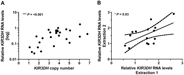
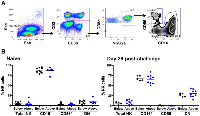
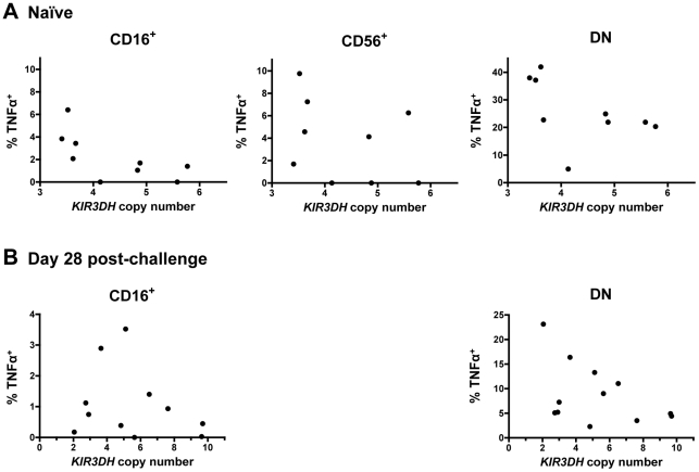
While the contribution of CD8⁺ cytotoxic T lymphocytes to early containment of HIV-1 spread is well established, a role for NK cells in controlling HIV-1 replication during primary infection has been uncertain. The highly polymorphic family of KIR molecules expressed on NK cells can inhibit or activate these effector cells and might therefore modulate their activity against HIV-1-infected cells. In the present study, we investigated copy number variation in KIR3DH loci encoding the only activating KIR receptor family in rhesus monkeys and its effect on simian immunodeficiency virus (SIV) replication during primary infection in rhesus monkeys. We observed an association between copy numbers of KIR3DH genes and control of SIV replication in Mamu-A*01⁻ rhesus monkeys that express restrictive TRIM5 alleles. These findings provide further evidence for an association between NK cells and the early containment of SIV replication, and underscore the potential importance of activating KIRs in stimulating NK cell responses to control SIV spread.


Conflict of interest statement

The authors have declared that no competing interests exist.

Figures

Figure 1
Figure 1. Intra-run reproducibility in KIR3DH copy number determination and validation of quantitative real-time PCR estimates of KIR3DH copy numbers by MLPA (multiplex ligation-dependent probe amplification).
(A) KIR3DH copy numbers were determined using triplicates of each DNA sample from 77 rhesus monkeys in two separate experiments to validate intra-experiment reproducibility (R2  =  0.87, β  =  0.816). (B) Comparison of signal ratios of two MLPA experiments. Pairs of ratios (20 samples) cluster around groups corresponding to KIR3DH copy numbers of 2, 3, 4 and 5. (C) Relationship between KIR3DH copy numbers determined by qPCR and MLPA (R2  =  0.77, β  =  0.729). The 95% confidence interval is shown by dashed lines.
Figure 2
Figure 2. Distribution of KIR3DH copy numbers in Indian-origin rhesus monkeys.
Copy numbers of KIR3DH genes were determined using quantitative real-time PCR on genomic DNA of 77 Indian-origin rhesus monkeys. (A) Distribution of KIR3DH copy numbers in the entire cohort of monkeys. (B) Boxplots of KIR3DH copy number distribution in rhesus monkeys divided into three cohorts: Mamu-A*01 and Mamu-A*01+ rhesus monkeys, Mamu-B*17 and Mamu-B*17+ rhesus monkeys and rhesus monkeys expressing only TRIM5 alleles 1–5 or expressing at least one TRIM5 allele from the group 6–11. (C) The median, mean and standard deviation (SD) of KIR3DH copy numbers are shown for various subgroups of rhesus monkeys. P values were calculated using the Mann-Whitney U test (two-tailed).
Figure 3
Figure 3. Association of KIR3DH copy numbers and peak plasma SIV RNA levels in Indian-origin rhesus monkeys.
KIR3DH copy numbers were determined by quantitative real-time PCR using genomic DNA from rhesus monkeys. Peak plasma SIV RNA levels were quantified on day 14 post-SIVmac251-infection. Scatter plots represent the relationship between SIV peak viral load and KIR3DH copy numbers in the entire cohort of rhesus monkeys (P  =  0.70) (A), in Mamu-A*01 + rhesus monkeys (P  =  0.24) (B) and in Mamu-A*01 rhesus monkeys (P  =  0.08) (C). The Mamu-A*01 + and Mamu-A*01 rhesus monkeys were subdivided into two groups: one group having KIR3DH copy numbers below the median (black) and the other group having KIR3DH copy numbers above the median (blue). These groups were further subdivided into monkeys expressing only TRIM5 alleles 1–5 or expressing at least one TRIM5 allele of the group 6–11. P values were determined using the Mann-Whitney U test (two-tailed).
Figure 4
Figure 4. Lack of association between KIR3DH copy numbers and clinical course following SIVmac251 infection in Mamu-A*01 rhesus monkeys.
Peripheral blood CD4+ T cell and central memory (CM) CD4+ T cell counts were measured by flow cytometry and complete blood counting. Loss of CD4+ T cells (A) and CM CD4+ T cells (B) were determined on days 14 and 70 post-challenge (pc). The Mamu-A*01 rhesus monkeys were divided into two groups: monkeys having KIR3DH copy numbers below (black) or above (blue) the median.
Figure 5
Figure 5. Expression of particular KIR3DH alleles is not associated with KIR3DH copy numbers.
KIR3DH cDNA clones of 8 unrelated rhesus monkeys with KIR3DH copy numbers ranging from 1–10 copies pdg were obtained using a PCR method described by Blokhuis et al. . Two previously reported KIR alleles (GU112262, GU112301) and ten novel KIR3DH alleles (JN613291-JN613300) were identified.
Figure 6
Figure 6. KIR3DH mRNA expression is associated with KIR3DH copy numbers in CD14CD16+ NK cells.
CD14CD16+ NK cells were isolated from PBMCs of naïve rhesus monkeys using immunomagnetic beads. RNA was extracted from these isolated cells and KIR3DH mRNA expression levels were determined by quantitative RT-PCR. Relative RNA expression was calculated using the 2−ΔΔCP method. (A) Scatter plots show the association between KIR3DH copy numbers and KIR3DH mRNA expression levels (P  =  <0.001, R2  =  0.51). (B) KIR3DH mRNA expression levels were measured a second time in the same animals after a one month interval (P  =  0.03, R2  =  0.33).
Figure 7
Figure 7. Distribution of peripheral blood NK cell subsets in Mamu-A*01 rhesus monkeys.
(A) Representative flow cytometric plots defining NK cells. Rhesus monkey NK cells were defined as CD3 CD8α+ NKG2A+, and CD16 and CD56 expression was used to delineate three primary NK cell subsets: CD16+, CD56+ and double negative (DN) NK cells. (B) Percentages of circulating total NK cells as well as CD16+, CD56+ and double-negative (DN) NK cells were compared in naïve rhesus monkeys and SIVmac251-infected rhesus monkeys on day 28 post-challenge. The rhesus monkeys were divided into animals having KIR3DH copy numbers below (black) or above (blue) the median.
Figure 8
Figure 8. Secretion of tumor necrosis factor α (TNFα) in NK cell subsets following stimulation with K562 cells.
PBMCs were stimulated with K562 cells at an effector-to-target-ratio of 10∶1. Percentages of TNFα+ cells above background in CD16+, CD56+ and DN NK cells were measured in peripheral blood of naïve rhesus monkeys (A) and SIVmac251-infected rhesus monkeys on day 28 post-challenge (B). For the day 28 post-challenge timepoint, insufficient CD56+ NK cells were acquired to allow an analysis.

References

    1. Biron CA. Initial and innate responses to viral infections--pattern setting in immunity or disease. Curr Opin Microbiol. 1999;2:374–381. - PubMed
    1. Cerwenka A, Lanier LL. Natural killer cells, viruses and cancer. Nat Rev Immunol. 2001;1:41–49. - PubMed
    1. Orange JS. Human natural killer cell deficiencies and susceptibility to infection. Microbes Infect. 2002;4:1545–1558. - PubMed
    1. Lanier LL. NK cell recognition. Annu Rev Immunol. 2005;23:225–274. - PubMed
    1. Trinchieri G. Biology of natural killer cells. Adv Immunol. 1989;47:187–376. - PMC - PubMed

Publication types

MeSH terms